Tag Archive for: Acetabulum Fractures

Iliofemoral Approach to Acetabulum

Vol 3 | Issue 1 | Jan – Apr 2017 | page:14-16 | Ravi Gupta, Ashwani Soni


Author: Ravi Gupta [1], Ashwani Soni [1]

[1] Department of Orthopaedics Government Medical College Hospital, Chandigarh 160031India.

Address of Correspondence
Dr Ravi Gupta
Department of Orthopaedics
Government Medical College Hospital, Chandigarh 160031
Email: ravikgupta@yahoo.com


Abstract

Surgical approach to acetabulum fractures depend on the type of fracture and fracture displacement along with surgical preference of the surgeon. Ilio-femoral approach is one of the most important approaches for acetabular fractures. It allows access to anterior column as well as anterior wall fractures. This symposium article details the surgical technique as well as indications and shortcomings of this approach.
Keywords: Ilio-femoral appraoch, acetabulum fractures, anterior column fractures.


Introduction

Operative reduction and fixation is considered as accepted treatment method for displaced acetabular fractures now days. Surgical approach to acetabulum may be anterior (ilioin-guinal, ilio-femoral, Stoppa approach [1,2]), posterior (Kocher-Langenbeck approach and variants [3]) or combined (simultaneous or different sittings) [4]. The choice of approach depends on the type of fracture, displacement and the surgeon’s preference [5].
Ilio-femoral approach provides wide access to the entire internal iliac fossa including visualization of the anterior aspect of the sacroiliac joint. This approach also provides digital and some visual access to the quadrilateral surface and greater sciatic notch. Medially, along the anterior column this approach provides access up to iliopectenial eminence.
Combined with Kocher-Langenbeck approach, anterior column component of T-type fracture can be fixed by this approach. When combined with pfannenstiel approach for symphysis pubis, almost all types of anterior column and anterior wall fractures can be addressed by this approach.
Surgical approach to acetabulum fractures depend on the type of fracture and fracture displacement along with surgical preference of the surgeon. Ilio-femoral approach is one of the most important approaches for acetabular fractures. It allows access to anterior column as well as anterior wall fractures. This symposium article details the surgical technique as well as indications and shortcomings of this approach
Anterior column fractures where main displacement is cephalad to the joint are the best candidates for this approach. Associated anterior + posterior hemitransverse fractures are also addressed by this approach.

Skin incision

Skin incision begins proximally posterior to the gluteus medius pillar, runs parallel to the iliac crest to the anterior superior iliac spine and then extend distally between sartoreus and tensor muscle. In modification to this approach, the dissection stops the anterior extent of proximal limb of skin incision 1-2 cm lateral to the anterior superior iliac spine and then extends distally and laterally directly over the anterior aspect of the tensor muscle belly. This reduces the damage to lateral cutaneous nerve and prevent to some extent the skin problems at the junction of two limbs of incision. Position of the proximal limb of incision need to be modified according to the body habitus of patient as scar directly over the iliac crest in a thin patient can be problematic.

Superficial dissection

Deeper dissection is done through subcutaneous tissue. External oblique muscle is released from the iliac crest leaving thick fascio/periosteal cuff for subsequent repair at the time of closure. This release of muscle is started posteriorly where it overhangs the iliac crest and extends anteriorly to the level of planned anterior superior iliac spine osteotomy or sartorius/ inguinal ligament release. Internal iliac fossa is exposed by lifting the iliac muscle subperiosteally and packed with sponge.

Osteotomy of ASIS

Osteotomy of ASIS is done with block measuring 2 cm in depth and 2 cm in anteroposterior diameter. Tensor attachment to this bony block is released. This osteotomised bone block along with its attachments of iliacus, external oblique, Sartorius, and inguinal ligamanet is displaced medially.
This osteotomy is not possible in cases where fracture line reaches to the area of osteotomy. In these cases Sartorius and inguinal ligament is released from ASIS as a single cuff of tissue.
Tensor-sartorius interval is identified and fascia of thigh is incised over tensor muscle belly starting 1-2 cm lateral to the ASIS. The tensor muscle is retracted laterally and the medial sheath of the tensor fascia is retracted medially protecting the lateral femoral cutaneous nerve. Subsequently the dissection is done through the floor of tensor sheath and the proximal and distal exposures and joined. Approximately 10 cm from the ASIS the ascending branches of the lateral femoral circumflex artery and vein are encountered under the aponeurotic fascial layer over the rectus femoris muscle which needs to be identified and ligated in case if wide exposure is required. True pelvis is accessed by releasing iliopectineal fascia from the pelvic brim starting just anterior to the SI joint and extending anteriorly to the level of the pubic root. subperiosteal elevation of the obturator internus from the quadrilateral surface can provide further access to pelvis.
At this stage entire iliac fossa and anterior sacroiliac joint is accessible. Quadrilateral lateral plate and greater sciatic notch is approached to certain extent either by direct visualisation or digital palpation. By retracting iliopsoas muscle medially, after relaxing it by doing flexion and adduction at hip joint; the exposure can be extended medially up to iliopectenial eminence.
Further access to anterior column, medial to iliopectineal eminence can be gained through a separate pfannenstiel incision for pubic symphysis. By doing this almost all types of fractures of anterior column can be addressed.
Corona mortise is an anastomosis behind the pubic symphysis between the obturator and external iliac arteries. By approaching medial most part of anterior column through pfannenstiel incision this anastomoses should be identified and ligated if present otherwise this anastamoses can be life threatening if injured.

Closure:
After placing the drain osteotomy of ASIS is fixed with 2.7 or 3.5 mm lag screws. External iliac muscle is sutured to iliac crest by fascial/periosteal repair. Fascial repair is done at distal limb of wound followed by subcutenous and skin closure.


How to Cite this article: Gupta R, Soni A. Iliofemoral Approach to Acetabulum. Trauma International Jan-Apr 2017;3(1): 14-16.
.


(Abstract)      (Full Text HTML)      (Download PDF)


History of Pelvi-Acetabular Fracture Treatment

Vol 2 | Issue 2 | May – Aug 2016 | page:17-19 | A S Prasad, Rahul Rishi


Author: A S Prasad [1], Rahul Rishi [1]

[1] Chandak Nursing Home Nagpur Maharashtra India.

Address of Correspondence
Dr. A S Prasad
Postal Address of correspondence with email of corresponding author-7/187 C ,Swaroop Nagar, Kanpur-208002.
E-mail: drprasadas@gmail.com


Abstract

Acetabular Fractures were treated conservatively before 1960. Robert Judet in 1960 started to treat displaced Acetabular Fractures Surgically. In the modern world of fast moving vehicular traffic, the Pelvic Fractures are the third most common cause of death in Motor Vehicle accidents. The associated injuries to the abdomen, chest, CNS and long bones as well as hemodynamic instability is frequently present Implementation of the ATLS protocol has helped in the early management of such patients but even during the ”golden hour” further decisions need to be taken regarding the surgical management (temporary or definitive) in order to improve survival rates in such patients. Acetabular Fixation can be delayed for a week after the index injury managing the life threatening injuries first. However they may have to be fixed as a damage control orthopedics when the Hemorrhage of pelvis fracture itself is a cause of the shock.
Keywords: Acetabulum Fractures, Pelvis Fracture, treatment.


Introduction

Today the Pelvic Fractures are the Third most common cause of death in Motor Vehicle accidents. The concern is to reduce this and guidelines for the management have come for the same. Historically too this has been discussed a lot. The modern specialty was really born with the publication of Malgaigne’s books on fractures and subluxations in 1847[2] . At the same time in the United Kingdom, Sir Astley Cooper (1768–1841) described various pelvic fractures, making the distinction between marginal stable fractures and unstable pelvic ring injuries. At this stage it should be emphasized that all these observations were made onclinical grounds with no x-rays. In the late 1950s and 1960s, Robert Judetbegan attempting to treat displaced acetabular fractures surgically.He felt that the outcomes with displaced acetabular fractures following conservative treatment were unacceptable. He identified certain subsets of acetabular fractures thatdid not do well with conservative treatment. These included fractures that involved the tectum or roof and those fractures where the hip was unstable. He described ten classic fractures patterns (five basic and five complex). Judet also developed many surgical approaches, particularly the ilio-inguinal approach, for treatment of anterior acetabular injuries.  The present day concept of fixation after appropriate reduction started with Judet. He emphasized that for pelvic fractures treated non-operatively functional outcome was frequently a problem for both patient and surgeon as deformities and chronic pain had a significant incidence.

Present Day Concepts

In the modern world with a fast moving life, the acetabular fractures are mostly a part of Poly Trauma. Prioritizing the steps of management is an issue to get the best for the injured. The associated injuries to the abdomen, chest, CNS and long bones as well as hemodynamic instability is frequently present Implementation of the ATLS protocol has helped in the early management of such patients but even during the ”golden hour” further decisions need to be taken regarding the surgical management (temporary or definitive) in order to improve survival rates in such patients[1]. Haemodynamic stabilization is of capital importance in unstable and ”in extremis” patients. Hemorrhagic shock is one of the four pathological cascades of polytrauma (shock, hypothermia, coagulopathy, soft tissue injury)[3].Focus is given on the identification of the source of bleeding (fracture site, thoracic, abdominal or pelvic organ injuries, wounds and arteriovenous disruptions). Blunt abdominal trauma, open fractures and wounds should be addressed promptly. Normo-voluemia can then be restored primarily by administration of crystalloids/colloids and blood products.

Damage Control Orthopedics in Fracture Acetabulum

In some patients who are both hemodynamically and mechanically unstable, and in whom the major bleeding is thought to be related to the pelvic fracture, external stabilization of the pelvis becomes the first priority. Because the main sources of bleeding are most frequently the presacral venous plexus and fractured bony surfaces. External stabilization decreases the hemorrhage by reducing the volume of the pelvic basin and approximating the fracture ends.

Pelvic binders
Circumferential pelvic binders or sheets are gradually replacing anterior external fixation (AEF) as the method of choice of immediate external stabilization, and currently form part of the ATLS protocol[4]. These binders are noninvasive, simple to apply, inexpensive and can be applied at a prehospital stage.
It has been shown that simple application of this sling increases pelvic stability by 61% in response to rotational stress and 55%, flexion–extension. Although the same study[5] found this method to be less rigid than AEF, it has nonetheless been shown to reduce unstable pelvic fractures radiologically and to improve patient’s hemodynamic status[6].
Clinical judgment and reassessment are important in using these techniques. This safe, noninvasive method seems to be a logical first resuscitative step with a serious pelvic fracture, to provide early hemorrhage control before considering invasive methods

Anterior external fixation
Immediate AEF of an unstable pelvic injury has been the mainstay of acute stabilization for the past few decades. Reimer and coworkers[7] reduced mortality rates from 22% to 8% by adding acute AEF to their hospital resuscitation protocol. Based on their results, they concluded that skeletal stabilization of pelvic injury should be viewed as a part of resuscitation rather than reconstruction. Burgess and colleagues[18] and others[9,10] have also documented decreased transfusion needs and reduced mortality with the use of anterior external fixator. Subsequent investigators [8,9,11,14] have also recommended immediate application of external fixation for hemodynamically unstable patients, and consider it a life-saving procedure. Some investigators have advised[9] prophylactic stabilization with anterior external fixator in all patients demonstrating bony instability, as even those patients who are initially hemodynamically stable on presentation may decompensate later. The anterior fixator is thought to contribute to hemostasis by maintaining a reduced pelvic volume, allowing tamponade, and by decreasing bony motion at the fracture site, allowing clots to stabilize[15]. C– clamps. To deal with posteriorly unstable fractures, Ganz and coauthors[16] developed a pelvic C-clamp, now available in most trauma units. It acts like a simple carpenter’s clamp and can exert transverse compression directly across the sacroiliac joint. Experimental data[16] have shown that an average compression force of 342 N can be applied to the area of this joint. These clamps have been used therapeutically in hemodynamically unstable patients, and prophylactically in stable patients with unstable pelvic- ring disruptions. Hemodynamic status and fracture reduction have been shown to improve in both groups[16].

Acute fracture fixation
Provisional fixation of unstable pelvic- ring disruptions with a pelvic clamp or an external frame with a supracondylar pin has proved markedly beneficial in the resuscitative phase of management. If the patient is too ill to allow a more invasive intervention, traction pins can remain in place with the external frame as definitive treatment. If, however, the patient undergoes a laparotomy to deal with visceral injuries, symphyseal disruption and medial ramus fractures should be plated at the same time. Because neither blood loss nor operative time is greatly increased, combining these repairs decreases the risk of complications in a patient who is already compromised[17].

Pelvic packing
Patients who remain unstable with a probable retroperitoneal cause in spite of aggressive resuscitative efforts should not be transported to a distant angiography suite, especially if delay is involved[18]. These are often patients at risk for abdominal compartment syndrome, and who therefore need an open peritoneal cavity for adequate cardiovascular physiologic support after surgery. Such patients undergoing laparotomy for an identifiable intraperitoneal cause of hemorrhage should be assessed for an expanding pelvic hematoma. The true pelvis should be packed at that time if the hematoma has ruptured; the pelvic hematoma is otherwise not opened routinely. The true pelvis should be packed with large abdominal swabs and the wound closed over the packs to create tamponade. The packs are removed or changed in a second procedure at 24–48 hours.

Open fractures

Potentially lethal injuries with a reported mortality rate of 30%–50%[19]. Open fractures of the pelvis by definition communicate with the rectum, the vagina, or the outside environment by disruption of the skin. They are often associated with disruption of the pelvic floor, leading to loss of tamponade and persistent bleeding. Clinical suspicion of an open fracture and any rectal or vaginal bleeding mandate a thorough examination, proctoscopic, sigmoidoscopic or by speculum.
These are the cases for application of Damage Control Orthopedics Principles.

Definitive Fixation
Since the birth of pelvic fracture surgery, timing of surgery has been referred to as either early or late. Unfortunately, the terminology of ”early/late fixation” has been used inconsistently. Some of them define as ”early” the first 8 h[20],24 h[21],72 h[22],first week[23] or even the first 14 days[22] or 21 days[24] post-injury and the term ”late” has been used for periods above 2 weeks post-injury[25] or as long as 3 months post-injury[26]. Definitive fixation in 1st week is advisable, however can be extended to 14 days is the majority opinion now.

Delayed Fixation
Even though the studies favoring Early Total Care became the golden rule in trauma surgery, there was still much controversy. Patients were operated between the 5th and 14th day based on the assumption that earlier operation would be associated with significant and possibly life- threatening hemorrhage[27] and hemodynamically unstable patients were generally considered to be too unstable to undergo even external fixation[28].In real life, delayed fixation can be due to medical complications, unstable patients unable to operate on or transfer to a specializedCentre, unavailable senior surgeon, unavailable operating staff and operating rooms.Delayed fracture fixation creates operative difficulties: scar tissue, callus formation, inability toobtain anatomic reduction and need for more extensile approaches. Mears et al. found thatdelayed surgery of more than 11 days was related tosignificantly fewer anatomical reductions[29]. Alsothe quality of reduction is strongly related to theage of the patient, the above 70 year-old havingpoorer reductions and more intra-articular damage[29].

Conclusion

Fractures around hip joint are one of the most common and debilitating entities requiring intervention . These fractures encompass fractures of the pelvis and acetabulum. Health care providers must be trained and educated with scope of this problem as well as the basic types of these fractures and treatment they warrant. The clear distinction between energy levels of the injuries leading to these fractures should be understood and treatment given accordingly. This article briefly amalgamates these issues and gives pictorial examples to illustrate the specific points.


References

1. Katsoulis E, Pape HC, Giannoudis PV. Shock-room management of pelvic ring lesions. Eur J Trauma 2005;31(3):222— 30.
2. Malgaigne JF (1847) Traite des fractures et des luxations,2 volumes.
3. Pohlemann T, Bosch U, Gansslen A, Tscherne H. The Hann- over experience in management of pelvic fractures. Clin Orthop Relat Res 1994;(305):69—80.
4. American College of Surgeons. Advanced Trauma Life Support for Doctors. Instruc- tor Course Manual. Chicago (IL): the College; 1997. p. 206-9.
5. Bottlang M, Krieg JC, Mohr M, Simpson TS, Madey SM. Emergent management of pelvic ring fractures with use of circum- ferencial compression. J Bone Joint Surg Am 2002;84(Suppl 2):43-7.
6. Simpson T, Krieg JC, Heuer F, Bottlang M. Stabilization of pelvic ring disruptions with a circumferencial sheet. J Trauma 2002;52:158-61.
7. Reimer BL, Butterfield SL, Diamond DL, Young JC, Raves JJ, Cottington E, et al. Acute mortality associated with in- juries to the pelvic ring: the role of early patient mobilization and external fixa- tion. J Trauma 1993;35:671-7.
8. Burgess AR, Eastridge BJ, Young JW, El- lison TS, Ellison PS Jr, Poka A, et al. Pelvic ring disruptions: effective classifica- tion system and treatment protocols. J Trauma 1990;30:845-56.
9. Poka A, Libby E. Indications and tech- niques for external fixation of the pelvis. Clin orthop 1996;(329):54-9.
10. Gylling SF, Ward RE, Holcroft JW, Bray TJ, Chapman MW. Immediate external fixation of unstable pelvic fractures. Am J Surg 1985;150(6):721-4.
11. Kellam JF. The role of external fixation in pelvic disruptions. Clin Orthop 1989; (241):66-82.
12. Slatis P, Karaharju EO. External fixation of unstable pelvic fractures: experience in 22 patients treated with trapezoid com- pression frame. Clin Orthop 1980;(151): 73-80.
13. Trafton PG. Pelvic ring injuries. Surg Clin North Am 1990;70:655-69.
14. Wild JJ Jr, Hansen JW, Tullos HS. Un- stable fractures of the pelvis treated by ex- ternal fixation. J Bone Joint Surg Am 1982;64:1010-20.
15. Mears DC. Clinical techniques in the pel- vis. In: Mears DC, editor. External skele- tal fixation. Baltimore (MD): Williams and Wilkins; 1983. p. 342.
16. Ganz R, Krushell AJ, Jakob RP, Kuffer J. The antishock pelvic clamp. Clin Orthop 1991;(267):71-8.
17. Tile M. Acute pelvic fractures: II. Princi- ples of management. J Am Acad Orthop Surg 1996;4(3):152-61.
18. Gansslen A, Giannoudis P, Pape HC. Hemorrhage in pelvic fractures: Who needs angiography? Curr Opin Crit Care 2003;9:515-23.
19. Jones AL, Powell JN, Kellam JF, McCor- mack RG, Dust W, Wimmer P. Open pel- vic fractures: a multicenter retrospective analysis. Orthop Clin North Am 1997;28 (3):345-50.
20. Latenser BA, Gentilello LM, Tarver AA, et al. Improved outcome with early fixation of skeletally unstable pelvic fractures. J Trauma 1991;31(1):28—31.
21. Plaisier BR, Meldon SW, Super DM, Malangoni MA. Improved
outcome after early fixation of acetabular fractures. Injury 2000;31(2):81—4.
22. Cole JD, Bolhofner BR. Acetabular fracture fixation via a modified Stoppa limited intrapelvic approach. Description of operative technique and preliminary treatment results. Clin Orthop Relat Res 1994;(305):112—23.
23. Browner BD, Cole JD, Graham JM, et al. Delayed posterior internal fixation of unstable pelvic fractures. J Trauma 1987;27(9):998—1006.
24. Matta JM, Tornetta III P. Internal fixation of unstable pelvic ring injuries. Clin Orthop Relat Res 1996;(329):129—40.
25. BruetonRN.Areviewof40acetabularfractures:theimpor- tance of early surgery. Injury 1993;24(3):171—4.
26. Johnson EE, Matta JM, Mast JW, Letournel E. Delayed reconstruction of acetabular fractures 21—120 days follow-
ing injury. Clin Orthop Relat Res 1994;(305):20—30.
27. Goldstein A, Phillips T, Sclafani SJ, et al. Early open reduc- tion and internal fixation of the disrupted pelvic ring. J Trauma 1986;26(4):325—33.
28. Riemer BL, Butterfield SL, Diamond DL, et al. Acute mor- tality associated with injuries to the pelvic ring: the role of early patient mobilization and external fixation. J Trauma 1993;35(5):671—5.
29. Mears DC, Velyvis JH, Chang CP. Displaced acetabular frac- tures managed operatively: indicators of outcome. Clin Orthop Relat Res 2003;(407):173—86.


How to Cite this article: Prasad AS, Rishi R. History of Pelvi-Acetabular Fracture Treatment. Trauma International May – Aug 2016;2(2):17-19.


(Abstract)      (Full Text HTML)      (Download PDF)


Emergency Management of Pelvic Fractures

Vol 2 | Issue 2 | May – Aug 2016 | page:25-30 | Nadeem Akhtar Faruqui


Author: Nadeem Akhtar Faruqui [1]

[1] Regency Hospital, Kanpur , Uttar Pradesh, India.

Address of Correspondence
Dr Nadeem A Faruqui
14/116d, Civil Lines,
Kanpur 208001 India
Email: nafaruqui@hotmail.com


Abstract

Hemorrhage is leading cause of death following pelvic fractures. 15-25% of patients of closed pelvic fractures and 50% of open pelvic fracture patients die as a result of haemorrhage. The main source of hemorrhage is the shearing injury of posterior thin walled venous plexus (80%). Other sources of bleeding include the bleeding cancellous bone and arterial injury. Injury to the pelvic viscera is also quite common because of the close proximity. Perineal lacerations make a closed injury into an open injury thereby more than doubling the risk of morbidity and mortality. Adequate fluid replacement and application of a pelvic Binder or Sheet can markedly reduce the mortality associated with this fracture. A linen sheet folded onto itself to make it about 20-30 cms wide applied tightly over the pelvis and centered over the trochanters can significantly reduce the mortality figures.
Keywords: Acetabulum fractures, pelvis fracture, emergency management.


Introduction

The basic principles of emergency management, Airway, breathing and circulation hold true for pelviacetabular fractures too. However there are certain practical tips and principles that will help in getting better results in these situations. These principles and tips are detailed below.

Hemorrhage is Leading Cause of Death
15-25% for closed pelvic fractures and 50% for open pelvic fractures are fatal secondary to exsanguination following either external or internal occult bleeding [1,2]. Increased mortality associated with
– systolic BP <90 on presentation
– age >60 years
– increased Injury Severity Score (ISS)
– need for transfusion > 4 units
– Head and neck injuries

Sources of Major Bleeding
• intrathoracic
• intraabdominal
• retroperitoneal
• extremity (thigh compartments)
• pelvic
• Common source of hemorrhage
• venous injury (80%)
• shearing injury of posterior thin walled venous plexus
• bleeding cancellous bone
• Less common sources of hemorrhage
• arterial injury (10-20%)
• superior gluteal most common (posterior ring injury, APC pattern)
• internal pudendal (anterior ring injury, LC pattern)
• obturator (LC pattern)

Blood Transfusion
Replacement volume is estimated by using the formula of 3 ml of crystalloid for each 1 mm of blood loss. A minimum of 2 litres of crystalloid solution is given rapidly if the patient is in shock. Crystalloid is administered until type-specific blood of non–cross-matched universal donor (O-negative) is available
• Packed RBC:FFP:Platelets should ideally be transfused in the ratio 1:1:1
• this ratio has been shown to decrease mortality in patients requiring massive transfusion

D) Disability
Clinical Examination
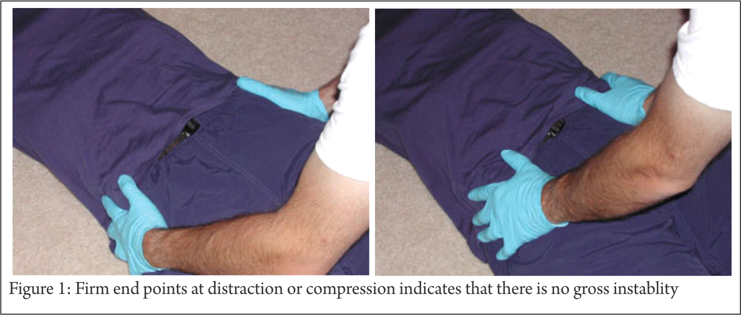
Pelvis: NO firm endpoint on rotation or traction indicates that there is no gross instablity (Fig 1). Pelvic compression and distraction test can detect gross instabilities, however these may at times be dangerous and can dislodge clots leading to further bleeding [3,4]. These test have been reported to have poor sensitivity and specificity and are no longer recommended in cases of pelvic fractures [4]

Perineum (Fig 2): Look for
• lacerations of perineum
• degloving injuries
• flank hematoma
• scrotal, labial or perineal hematoma, swelling or ecchymosis
• urethral bleeding

Rectal Examination
mandatory to rule out occult open fracture
• rectal blood
• anal sphincter tone
• bowel wall integrity
• bony fragments
• prostate position

Vaginal Examination
mandatory to rule out occult open fracture
Presence of blood in vaginal vault
Vaginal lacerations

Urogenital Examination (Fig 3)
Bladder and the urethra are most frequently injured (25-30% of major pelvic fractures). 66% male versus 34% females patients difficulty in voiding urine or blood at the urethral meatus.
95% of bladder injuries have gross haematuria
urethral injuries are suspected under following clinical scenario
• blood at the urethral meatus
• gross hematuria
• inability to spontaneously void
• high riding prostate on rectal examination
• retrograde urethrogram (RUG) should ideally be done before insertion of Urinary catheter in unstable patient with suspected urethral injury make only 1 attempt to pass the urinary catheter, if it fails do RUG

Neurologic Examination
• rule out lumbosacral plexus injuries (L5 and S1 are most common)
• rectal exam to evaluate sphincter tone and perirectal sensation

Whenever an unstable pelvic injury is suspected in a haemodynamically unstable patient, a pelvic binder/sheet should be applied to control bleeding
The goal of treatment for pelvic fracture stabilization is early control of life-threatening hemorrhage. Returning pelvic bones to correct position helps to reduce pelvic volume and control venous bleeding.

Pelvic Binder [5,6]
• Applies compression leaving less space for blood to accumulate. It decreases the pelvic volume and also reduces pelvic fractures
• Tamponades bleeding sources, such as fractured bony surfaces or ruptured vessels
• Reduces instability of the injured pelvis
• Prevents further damage to pelvic organs and vessels
• Reduces pain by limiting movement of pelvis
Commercially available pelvic binder are also useful but use of lumbosacral belts is to discouraged [6].

Pelvic Binder/sheet
Indications
• initial management of an unstable pelvic ring injury
Contraindications
• hypothetical risk of over-rotation of hemipelvis and hollow viscus injury (bladder) in pelvic fractures with internal rotation component (LC)
• no clinical evidence exists of this complication occurring
Technique
• A linen sheet folded onto itself to make it about 20-30 cms wide is passed under the pelvis
• Centered Over Greater Trochanters to effect indirect reduction
• Applied very tightly over the pelvis
• do not place over iliac crest/abdomen as it makes the ineffective and precludes assessment of abdomen
• augmented with traction & internal rotation of lower extremities and taping at knees & ankles
• transition to alternative fixation as soon as possible
• prolonged pressure (>24 hours) from binder or sheet may cause skin necrosis
• working portals may be cut in sheet to place percutaneous fixation
Lumbosacral belt should not be used because it is elastic and cover abdomen.
Important note. Binder should be centered over the TROCHANTERS and Not the ASIS

Because of their ease of use and fast application, Pelvic Binders have
largely replaced the Pelvic C-Clamp and External Fixators for early
mechanical stability in pelvic fracture [5].

Radiology & CT

X-ray Pelvis –AP
Pelvis Is Unstable When There Is
Sacro-Iliac joint Diastasis > 1 cm or
Cephalad Displacement of Posterior Sacro-Iliac Complex > 1 cm
Usually associated with Avulsion fracture of ischial spine, ischial tuberosity, sacrum or transverse process of 5th lumbar vertebrae

High Risk Patients as per the Young and Burgess Classification:
– LC III
– APC II
– APC III
– VS
– CM

Ct Scan is the Gold Standard

CT scan is essential in determining:
Posterior ring instability
Helps define comminution and fragment rotation
Intra-articular fragments
Fractures of articular surface of acetabulum and femoral head

Ultrasonography
Allows Focused Assessment with Sonography for Trauma (FAST)
Four classic areas are examined quickly by ultrasonography for free fluid (Blood):
Perihepatic space (hepatorenal recess)
Perisplenic space
Pericardium
Pelvis

External Fixation/ Stabilization of Pelvic Ring Injury
These are temporary life saving measures in the acute stage to stabilize the pelvis in a haemodynamically unstable patient. However, None of these methods can fully stabilize the pelvis. These are put on till such time as more definitive fixation can be done.
Femoral traction pin with 10-12 kg weight should be applied to maintain the reduction of the hemipelvis.

Mode of Action of External Fixation
•Decreases pelvic volume
•Stabilizes bleeding bone surfaces and venous plexus in order to form clot
•Reduces Pain

Types
A) Pelvic Binder/sheet
B) Pelvic C Clamp
C) External Fixator

A) Pelvic Binder/Sheet
Easily available everywhere annd is easy to apply. Technique of application discussed previously

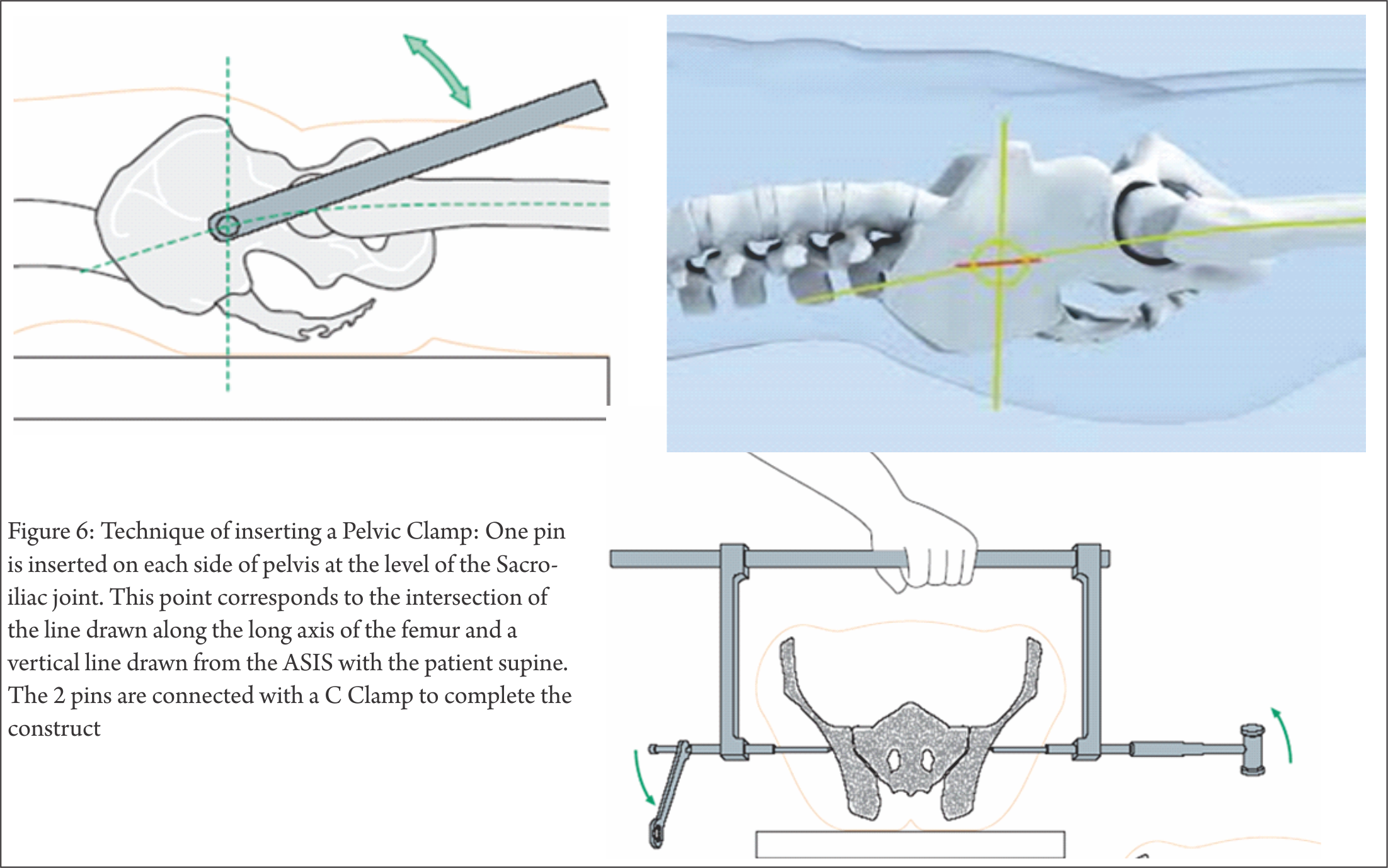
B) Pelvic C Clamp (Fig 6)
Indications
– Emergency stabilization of Sacroiliac joint disruptions and fractures of the sacrum with associated circulatory instability
Contraindications
Absolute contraindications are:
•Fracture lines within the illium (transiliac fracture) as it bears the risk of pin perforation through the fracture line
•Hemodynamic stability in Pelvic fractures
Relative contraindications are:
•Hemodynamic stability of the patient after Unstable type injuries
•Comminuted sacral fractures with risk of compression of the sacral nerve plexus

In life threatening situations hemorrhage control takes priority over the potential risk of nerve root compression
One Pin is inserted on each side of pelvis at the level of the Sacro-iliac joint. This point corresponds to the intersection of the line drawn along the long axis of the femur and a vertical line drawn from the ASIS with the patient supine. The 2 pins are connected with a C Clamp.
Advantage
Can be applied in the emergency room
Laparotomy can be done with the C Clamp in place
Disadvantage
Costly and not available everywhere.
Efficacy similar to Pelvic Binder/ Sheet.

C) External Fixator
Indications
Pelvic ring injuries with an external rotation component (apc, vs, cm)
Provide only marginal stability in vertically unstable ring injury

APC II (OPEN BOOK injuries with posterior ligaments/hinge intact) :
 All designs work
APC III injuries (Post Ligaments damaged)
 No designs work well (but AIIS frames better than ASIS frames)
Contraindications
•Ilium fracture that precludes safe application
•Acetabular fracture

Technique
Pin insertion in iliac crest (ASIS) (Fig 7)
multiple half pins inserted in the superior iliac crest placed in thickest portion of anterior ilium or gluteus medius tubercle

Should be placed before emergent laparotomy
• Stab incision over iliac crest
• Pass 2 K-wires by hand 1 each on medial & lateral sides of iliac wing
• Drill hole started at junction of Medial 1/3 & lateral 2/3rd of iliac crest
• 45 degree inclination lateral to medial
• Ceplalad to caudal direction – towards acetabulum
• Drill 1 cm
• 5 mm Schanz pin inserted with hand
• Cortical walls to guide pin into position
• Obturator Oblique view on C-Arm /Outlet view

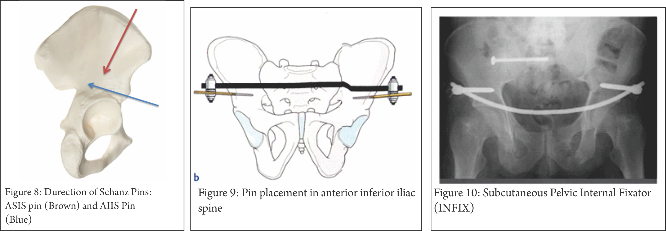
Pin Placement In Anterior Inferior Iliac Spine (AIIS) Fig 8,9)
single pin in column of supra-acetabular bone from AIIS towards PSIS. Obturator outlet or “teepee” view can be used to visualize this column of bone. AIIS pins can place the lateral femoral cutaneous nerve at risk
Tranverse skin incision at or below AIIS
•Muscles split longitudinally to avoid lateral femoral cutaneous nerve injury
•Drill through trocar under image intensifier towards greater sciatic notch
•Pins directed 30-450 towards midline in frontal plane
•Pins directed perpendicular to body axis or slightly cephalad
•Schanz pins of 50-70mm thread length required
•Fixator bar connects the pins on both sides
•Permits easy access for laparotomy

Subcutaneous Pelvic Internal Fixator (INFIX) (Fig 10)

One pedicle screw is fixed in the supra-acetabular bone of the ilium on each side. The pedicle screws are connected to each other by a rigid, anteriorly bowed fixation rod passed subcutaneously superficial to the sartorius muscles [7]
Less infection and wound site morbidity

After Stabilization/ External Fixation Of Pelvic Ring Injury
A) Control Hemorrhage
B) Control Contamination

A) Control of Haemorrhage

1) Pelvic Packing (Fig 11)
Make Midline Incision, pack the wound with sponges. Apply External Fixator. DON’T TRY TO LIGATE THE BLOOD VESSELS..Re-open wound after 24-48 hours and then ligate the vessels if possible or required.

2) Angiography

Indications
•Small bore artery (sup gluteal or obturator) can be controlled by embolization
•No role in Venous or bony bleeding
Available only in select centres and success based on multiple variables including: stability of patient, proximity of angiography suite, availability and experience of staff
CT angiography useful for determining presence or absence of ongoing arterial hemorrhage (98-100% negative predictive value)

Contraindications
• not clearly defined
Technique
•selective embolization of identifiable bleeding sources
• if uncontrolled bleeding even after selective embolization, bilateral temporary internal iliac embolization may be effective
• complications include gluteal necrosis and impotence

B) Control of Contamination
• Debridement & Packing of Open Wounds
• Suprapubic Catherization
• Diverting Colostomy

Conclusion

Priciples of pelvis fracture damage control
External Fixation
Pelvic Binder
Pelvic C-clamp
External Fixator
Control of Haemorrhage
Pelvic packing
Angiography

Control of Contamination
Debridement & packing of Open Wounds
Suprapubic catherization
Diverting colostomy.


References

1. O’Sullivan RE, White TO, Keating JF. Major pelvic fractures: identification of patients at high risk. J Bone Joint Surg Br. 2005;87(4):530-3.
2. Kido A, Inoue F, Takakura Y, Hoshida T. Statistical analysis of fatal bleeding pelvic fracture patients with severe associated injuries. J Orthop Sci. 2008;13(1):21-4.
3. Lee C, Porter K. The prehospital management of pelvic fractures. Emerg Med J. 2007 Feb;24(2):130-3
4. Grant PT. The diagnosis of pelvic fractures by ‘springing’. Arch Emerg Med. 1990 Sep;7(3):178-82.
5. Bottlang M, Krieg JC, Mohr M, Simpson TS, Madey SM. Emergent management of pelvic ring fractures with use of circumferential compression. J Bone Joint Surg Am. 2002;84-A Suppl 2:43-7
6. Mohanty K, Musso D, Powell JN, Kortbeek JB, Kirkpatrick AW. Emergent management of pelvic ring injuries: an update. Can J Surg. 2005 Feb;48(1):49-56.
7. Vaidya R, Kubiak EN, Bergin PF, Dombroski DG, Critchlow RJ, Sethi A, Starr AJ. Complications of anterior subcutaneous internal fixation for unstable pelvis fractures: a multicenter study. Clin Orthop Relat Res. 2012 Aug;470(8):2124-31.


How to Cite this article: Faruqui NA. Emergency Management of Pelvic Fractures. Trauma International May-Aug 2016;2(2):25-30.


(Abstract)      (Full Text HTML)      (Download PDF)


Management of Acetabulum Fractures – Basic Principles and Tips and Tricks

Vol 2 | Issue 2 | May – Aug 2016 | page:20-24 | Atul Patil, Ashok Shyam, Parag K Sancheti


Author: Atul Patil [1], Ashok Shyam [1], Parag K Sancheti [1]

[1] Sancheti Institute for Orthopaedic and Rehabilitation, Pune. Maharashtra, India.

Address of Correspondence
Dr. Ashok Shyam,
Sancheti Institute for Orthopaedic and Rehabilitation, Pune. Maharashtra, India.
E-mail address: drashokshyam@gmail.com


Abstract

Acetabulum fractures require systematic approach for understanding the fracture pattern and also for planning the treatment plan. The fractures have to be correctly identified radiologically and clear definition of fracture patterns should be made before planning. The radiological parameters must be kept in mind in planning of surgical approach and also the fixation method. This may require a long learning curve but these basics have to be kept in mind while dealing with acetabulum fractures. There are new techniques like 3d CT, virtual assessment of the fracture, 3D print modelling of the fractures that may help in complex fractures, but the basic principles remain the same. Advancements in technology simply refines the ways and means of interpretation and implementation of the basic principles. The current article is compilation of experience gathered over a period of time. The entire article emphasizes on the basics of understanding and managing acetabular fractures and also includes important tips and tricks that facilitate the treatment.
Keywords: Acetabulum fractures, surgical management, Letournal and judet.


Introduction

Acetabular fractures are still difficult fractures to manage and are a major challenge to treating orthopaedic surgeon [1]. Pioneering work was done by Letournal and Judet in 1964 [2]. They systematically classified acetabular fractures and developed a logical line of thinking for dealing with these fractures. They improved the understanding of morphology and popularized surgical principles for management of these injuries. Letournal and Judet put forth the two column theory of acetabulum anatomy. They envisioned acetabulum to be made of two columns. Anterior column from below the sacroiliac joint to the ischial tuberosity and posterior column from superior iliac crest to pubic symphysis with both columns attached to the sacrum by thick strut of bone lying above greater sciatic notch and called sciatic buttress [Fig. 1].

Based on these anatomical factors they suggested the first systematic classification of acetabular fractures.2 Although comprehensive classification is necessary for investigational purposes such as prognosis and outcome studies, it is less important in making decisions on individual cases. Every acetabulum fracture case is different, therefore, trying to force square plug in a round hole is  counterproductive. The surgeon must know the basic fracture types, but even more important, he must be able to interpret the radiographs and draw the fracture lines on a dry skeleton. The 3d CT virtual model and 3D print life size models of fractures acetabulum also need the basic understanding of the fracture anatomy and are helpful only when such clear understanding is present. This clarity also helps in selecting the surgical approach. Most of the innovative work was performed by Letournal and Judet and their recommendation is still valid till date [2,3]. Anatomical reduction of the articular fragment and restoration of a congruent and stable hip are the two most important factors in management of acetabular fractures. Fractures reduced to less than 1 mm of articular step have less incidence of posttraumatic hip arthrosis and a better and long lasting functional restoration as compared to fractures reduced with 1 – 3 mm residual articular displacement [4,5].

To meet these goals congruent and stable hip joint, four objectives are to be kept in mind
1. Correct interpretation of the radiographs
2. Identification and understanding the fracture pattern
3. Choosing the appropriate management
4. Striving for best surgical result.

1. Correct interpretation of radiographs-

On the antero-posterior pelvis radiograph, six lines are identified: the ilioischial line, iliopectineal line, the weight bearing dome (sourcil), teardrop, anterior rim (acetabulo-obturator line), and posterior rim (ischioacetabular line) [Fig. 2].

The iliopectineal line represents the anterior column. The ilioischial line is equated with the posterior column but is not actually created by the posterior border of the innominate bone but by the cortex of the quadrilateral surface. Thus fractures that disrupt the quadrilateral plate are seen as discontinuity of the ilioischial line even though these fractures do not disrupt the posterior border. The radiographic roof represents the cranial portion of the acetabular articular surface namely the weight bearing dome of the acetabulum. The lateral limb of the teardrop represents the floor of the cotyloid fossa while the medial limb represents the lateral wall of the obturator canal. Splitting of tear drop is seen when fracture line transverses through these areas. The anterior and posterior rims give some idea about the wall fractures however they are better diagnosed on Judet views.
Obturator view- is taken with injured side up and pelvis tilted 45 degrees. The posterior column and the anterior wall are visualized well (Figure 3a).
Iliac view – taken with pelvis tilted 45 degrees and injured side down. The posterior column and the anterior wall are visualized well (Figure 3b).

2. Identifying and understanding the fracture pattern-

According to Brander and Marsh [6], answers to following eight questions about the radiographic observations are used to determine the acetabular fracture pattern:
A) Is a fracture of the obturator ring present? If the obturator ring is broken then the fracture is either a column type of’ fracture or a T-shaped fracture.
B) Is the ilioischial line disrupted? Disruption of the ilioischial line occurs in fractures involving the posterior column or fractures in the transverse group.
C) Is the iliopectineal line disrupted? Disruption of the iliopectineal line indicates anterior column involvement or one of the transverse-type fractures.
D) Is the iliac wing above the acetabulum fractured? Iliac wing fractures are observed in fractures involving the anterior column, anterior column with posterior hemitransverse or both column fractures.
E) Is the posterior wall fractured? Posterior wall fractures may occur in isolation or in combination with posterior column or transverse fractures.
F) Does the fracture divide acetabulum into front and back halves or front and bottom halves? T type fracture divides pelvis into top and bottom halves while a column type divides pelvis into front and back halves
G) Is the spur sign present? The spur sign is observed exclusively in the both-column fractures. The spur is a strut of bone extending from the sacroiliac joint. Usually, this strut of bone connects to the articular surface of the acetabulum. In the both-column fracture, this connection is disrupted; a fractured piece of bone that resembles a spur remains. The spur sign is best depicted on the obturator oblique view [Fig 4]
H) What is the orientation of major fracture line on CT scan?
According to the answers of these eight questions, the fractures can be classified using Letournal and Judet classification as shown in Table 1

3. Choosing the appropriate management pathway:

Need for surgical intervention can be determined by following two criteria’s
Fracture criteria’s – Unstable hip [the femoral head and acetabulum are non congruent on AP radiograph], Roof arc angle is less than 45°, Intraarticular fragments, Marginal impaction, Unreduced fracture dislocation
Patient factors – Age [>50 yrs think of conservative treatment and later date Total Hip arthroplasty when arthritis develops], Severe co morbidities [ASA grade III or more – Cx], pre existing hip arthritis [Cx and THA later], severe osteopenia, patients with psychiatric disorders, patients with restricted pre injury mobility.
First decide whether radiograph will require surgery, and then assess the patient for feasibility of surgical intervention. If answer to any of the above question is negative the fracture is treated conservatively.
Few Tips in patient assessment –
Morel-Lavalle´lesions contain liquefied hematoma and have been known to be culture positive nearly 30-50% of times. In such cases drain the hematoma and perform delayed surgery.
Complete neurological examination and documentation is necessary especially in posterior dislocation as it is associated with high incidence of sciatic nerve injuries [20%] which if discovered later gives unsatisfactory result to the patient and may lead to legal issues.
In case the surgery is delayed, skeletal traction is essential

4. Striving for best surgical result.

This involves a definite learning curve. Surgical approach is determined based on the fracture classification. There are four main approaches used for acetabular fractures viz.
A. Kocher-Langenbeck: Posterior wall, Posterior column, Transverse, Transverse PW, Posterior column PW, T shaped [Fig 5, 6].
B. Ilioinguinal: Anterior wall, Anterior column, Anterior Posterior Hemitransverse, Both- column fractures, Transverse (rare) [Fig 7].
C. Extensile iliofemoral approach: Both-column fractures, T shaped, Transverse PW, Fractures > 3 weeks involving both columns [Fig 8]
D. Combined: A single approach is always preferred however combined approaches may be needed for more complex fractures involving both columns.

4. Striving for anatomical reduction.

This is by far the most important variable affecting the outcome of acetabular surgery along with severity of initial trauma. It has a long learning curve and this aspect is highlighted by Matta and Merritt in their study of their first 100 acetabular fracture fixation cases [7]. They grouped the surgical reductions chronologically in groups of 20 consecutive patients and clearly established that with increased experience the ability to achieve anatomical reduction improved along with ability to avoid unsatisfactory results.

Tips and pearls for acetabular surgery

§ Keep three points in mind – Avoid Devascularization of Fragments, remove all intra-articular fragments, and try to achieve anatomical reduction.
§ After exposure, open and clean the fracture site and get intraarticular visibility by a wide capsulotomy which will help in assessing the intraarticular reduction. Keep a low throeshold for widening the exposure
§ Special instruments in form of reduction clamps etc must be kept ready and used when necessary to hold reduction and achieve provisional K wire fixation
§ Reduction of the fragments – this will require two things – traction to the femur and opening through the fracture.
-Traction can be applied by a traction table or direct traction via a corkscrew through femoral neck or a hook on greater trochanter might work as well.
– Open the fracture by removing the major piece out of the way and appreciate the impacted fragments. These fragments have to be reduced to achieve best result.
– In cases where there is a major posterior fragment [high transverse and major T – type], a Schantz pin with a T-handle can be introduced into the ischial tuberosity to manipulate the reduction.
§ Provide stable fixation – most reliable fixation is a lag screw compression. Achieve reduction of the fracture fragments and provisionally fix them with K wires. The fragment can be predrilled first, then reduced and held with two 1.6-mm smooth Kirschner wires. Then each wire is then sequentially replaced by lag screws. This method will prevent shift/toggle of the fracture fragment while insertion of the lag screws.
§ It is desirable to have two points of fixation for each fragment, however this may not be possible because of small size although use of mini screws may be considered
§ After this a neutralization plate is applied to augment the fixation. Here one should keep in mind that lag screws should always be placed along the rim of posterior wall fragments, and care should be taken to ensure that the plate buttressing the posterior wall are positioned as lateral as possible. Applying the buttress plate too medially, especially without rim lag-screw fixation, might result in loss of stabilization of the posterior wall
§ Keep in mind two points while fixing the fractures -Avoid over-contouring of the plate, put in more lag screws rather than a bigger plate
§ In cases with bi-columnar fractures the anterior fragment is fixed with lag screw in first stage. While reducing the posterior column sometimes the anterior column screw needs to be backed out to help get the best reduction after which the screw is re-tightened.

A word of caution about the posterior approach:
The sciatic nerve must be identified and protected by knee flexion. Muscle belly of short rotators should be used to protect the nerve while retraction. Sciatic nerve may vary with respect to its relationship with pyriformis but always lies behind the quadratus muscle and is best identified by this relationship.
. Superior gluteal artery and nerve lie in the greater sciatic notch in close relationship with the bone. They can be injured while stripping of the the periosteum and can retract into the pelvis where they can bleed profusely and are difficult to handle.
. Retraction of the hip abductors might be required for visualization of superior acetabulum; however this may cause traction injury to the superior gluteal nerve which supplies the major hip abductors and the gluteus medius and minimus muscles.
Risk of iatrogenic osteonecrosis of the posterior wall fracture fragments is caused by excessive stripping of their soft-tissue attachments. Every attempt should be made to maintain the capsular attachments to these posterior wall fragments.

Recent Advances

Rapid prototyping and 3D printing are fast coming up as refined diagnostic and planning tool for acetabulum fractures [8,9]. These techniques help in visuospatial visualization of fracture fragments and also determine the best approach and fixation methods and implants. Trajectories of the lag screws can be determined on the virtual 3D models and the same can be utilized during surgery. However the role will be limited to more complex fractures and further refinement of the procedure will help in establishing its role in definitive management of acetabular fractures.


References

1. Tile M, Helfet D, Kellam J. Fractures of the Pelvis and Acetabulum. Baltimore. Lippincott Williams & Wilkins; 3rd edition, 2003.
2. Judet R, Judet J, Letournel E. Fractures of the acetabulum: Classification and surgical approaches for open reduction. J Bone Joint Surg. 1964;46A:1615-38.
3. Letournel E. Fractures of the acetabulum. A study of a series of 75 cases. 1961. Clin Orthop Relat Res 1994;(305):5-9.
4. Letournel E, Judet R. Fractures of the acetabulum, 2nd ed. Berlin: Springer-Verlag, 1993.
5. Matta JM. Fractures of the acetabulum: accuracy of reduction and clinical results in patients managed operatively within three weeks after the injury. J Bone Joint Surg Am 1996;78(11):1632-45.
6. Brandser E, Marsh JL. Acetabular fractures: easier classification with a systematic approach. AJR Am J Roentgenol. Nov 1998;171(5):1217-28.
7. Matta JM, Merritt PO: Displaced acetabular fractures, Clin Orthop Relat Res 230:83, 1988.
8. Zeng C, Xing W, Wu Z, Huang H, Huang W. A combination of three-dimensional printing and computer-assisted virtual surgical procedure for preoperative planning of acetabular fracture reduction. Injury. 2016 Oct;47(10):2223-2227.
9. Duncan JM, Nahas S, Akhtar K, Daurka J. The Use of a 3D Printer in Pre-operative Planning for a Patient Requiring Acetabular Reconstructive Surgery. J Orthop Case Rep. 2015 Jan-Mar;5(1):23-5.


How to Cite this article: Patil A, Shyam AK, Sancheti PK. Management of Acetabulum Fractures – Basic Principles and Tips and Tricks. Trauma International May-Aug 2016;2(2):20-24.


(Abstract)      (Full Text HTML)      (Download PDF)