Tag Archive for: Intertrochanteric fractures

Classifications of Intertrochanteric fractures and their Clinical Importance

Vol 1 | Issue 1 | July – Sep 2015 | page: 7-11 | Dhiraj V Sonawane[1].


Author: Dhiraj V Sonawane[1].

[1] Grant Medical College and Sir JJ Group of Hospitals, Mumbai. India.

Address of Correspondence
Dr. Dhiraj V. Sonawane
Asst. Prof. Grant Medical College and Sir JJ Group of Hospitals, Mumbai.
Email: dvsortho@gmail.com


Abstract

Intertrochanteric fractures are one of the most common fractures encountered by an orthopaedic Surgeon. Many attempts to classify these fractures are made and different scientific rationale are applied by various authors. Here we tried to provide an overview of both old and new classification of intertrochanteric fractures and also provide with the clinical significance of the same
Keywords: intertrochanteric fractures, hip fractures, classifications


Introduction

Intertrochanteric (IT) fractures are most common fractures seen in elderly osteoporotic, usually due to simple fall in the house. With increasing number of elderly patients its number is estimated to be double by 2040 [1]. Understanding important factors in management of IT fracture like stability, reduction, role of posteriomedial wall, lateral wall, will help in choosing implant for better outcome. Most classifications are based on these factors and help in selecting management protocols. Many classification systems have come from last 6 decades, but none of them are found to be unanimously acceptable worldwide. Few classifications have focussed on stability and anatomical pattern (Evans; Ramadier; Decoulx; & Lavarde) while others on maintaining reduction of various types (Jensen’s modification of Evan’s, Ender; Tronzo, AO).
An ideal classification should be simple, reproducible, easy to apply and should provide information on stability after reduction, secondary displacement, technique of fixation, postoperative mobilisation, outcome, and also data organisation for research. It should have good interrater and intrarater reliability and validity.
Classification Review:
Various classifications in Intertrochanteric fractures:
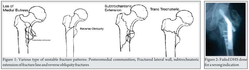
Evans Classification [2] (Fig 1):Fig 1 2 3
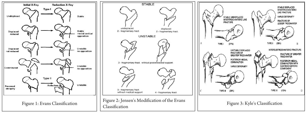
In 1949, Evans published his classification on intertrochanteric (IT) fractures as follows:
Type I:
Stable:
-Undisplaced fractures.
-Displaced but after reduction overlap of the medial cortical buttress make the fracture stable.
Unstable:
-Displaced and the medial cortical buttress is not restored by reduction of fracture.
-Displaced and comminuted fractures in which the medial cortical buttress is not restored by reduction of the fracture.
Type II: Reverse obliquity fractures.

Clinical importance: This helped in better understanding of intertrochanteric fractures based on stability of fracture after close reduction and skeletal traction. According to Evans, posterior-medial cortex continuation is important for restoring stability of IT fractures. Based on this he classified IT fractures into Stable and Unstable fractures. Stable fractures have intact or minimally communited posteriomedial cortex, while Unstable fracture has greater communition of posteriomedial cortex. Unstable fractures after reduction can be converted to stable fracture if the posteriomedial cortex opposition can be achieved. Reverse oblique pattern was considered inheritably unstable fracture as distal femur has tendency to drift medially due adductor pull.

Jensen’s Modification of the Evans Classification [3] (Fig. 2):
Jansen (1975 ) later modified Evans classification into three groups.
Displaced or undisplaced stable 2-fragment fractures, Unstable 3-fragment fractures with greater or lesser trochanter fracture and 4-fragment fractures

Clinical Importance: the classification reduced the number of types from 6 to 5 by including the extremely rare fracture with a reversed oblique fracture line and large greater trochanter fragment into Type 3. Modification of the Evans system offers the best prediction of the possibility of obtaining reliable anatomical reduction and the risk of secondary fracture dislocation.
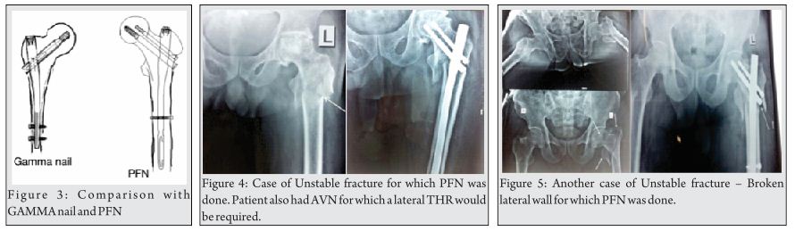
Kyle’s Classification [4] (Fig. 3):

Type I fractures consist of nondisplaced stable intertrochanteric fractures without comminution.
Type II fractures represent stable, minimally comminuted but displaced fractures; these are the fractures that, once reduced, allow a stable construct. Stable fractures are not a problem and hold up well with any type of fixation device.
Type III intertrochanteric fracture is a problem fracture and has a large posteromedial comminuted area.
Type IV fracture is uncommon and consists of an intertrochanteric fracture with a subtrochanteric component. This is the most difficult type of fracture to fix because of the great forces imposed by muscle forces and weight bearing on the subtrochanteric region of the femur.
Clinical Importance: Addition of new variant (type 4) extension of intertrochanteric fracture in neck.

AO/ Orthopaedic Trauma Association (OTA) Alphanumeric Classification [5] (1980-1987) (Fig. 4):Fig 4
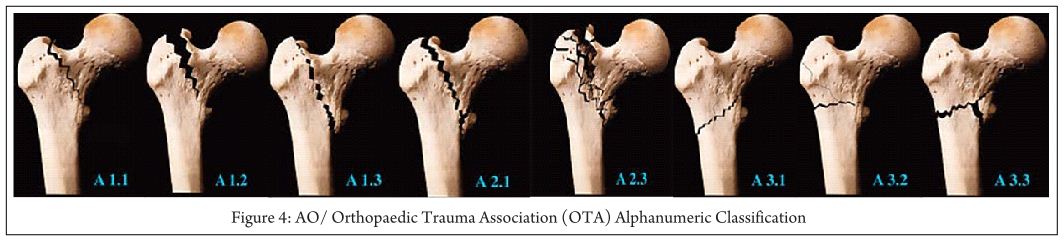
In the Comprehensive Classification of Fractures of the Long Bones, Müller and colleagues coded proximal hip fractures to offer a uniform alphanumeric fracture classification. This system was advocated by the AO/ASIF, and later adopted by OTA in their Fracture Compendium.
According to AO/OTA alphanumeric classification intertrochanteric fractures (Type 31A) Bone = femur = 3,Segment = proximal = 1,Type = A1, A2, A3 A1: simple (two-part) fractures, with the typical oblique fracture line extending from the greater trochanter to the medial cortex; the lateral cortex of the greater trochanter remains intact.
A2: fractures are comminuted with a posteromedial fragment; the lateral cortex of the greater trochanter, however, remains intact. Fractures in this group are generally unstable, depending on the size of the medial fragment
A3: fractures are those in which the fracture line extends across both the medial and lateral cortices; this group includes the reverse obliquity pattern or subtrochanteric extensions.
31-A Femur, proximal trochanteric
31-A1 Peritrochanteric simple
31-A1.1 Along intertrochanteric line
31-A1.2 Through greater trochanter
31-A1.3 Below lesser trochanter
31-A2 Peritrochanteric multifragmentary
31-A2.1 With one intermediate fragment
31-A2.2 With several intermediate fragments
31-A2.3 Extending more than 1 cm below lesser trochanter
31-A3 Intertrochanteric
31-A3.1 Simple oblique
31-A3.2 Simple transverse
31-A3.3 Multifragmentary.
Clinical importance: This helps in predicting prognosis and suggests treatment for the entire spectrum of IT fractures. Fractures A1.1 through A2.1 are commonly described as stable, and fractures A2.2 through A3.3 usually are unstable.
Generally, the Evans-Jensen type I fracture is represented by the 31-A1 group. Evans-Jensen type II fractures are in the 31-A2 group. The so-called reverse obliquity intertrochanteric fracture is in group 31-A3. It’s alphanumeric and standardized format make this system useful, particularly for research and documentation.

Boyd and Griffin Classification (1949) [6] (Fig. 5): Fig 5 6
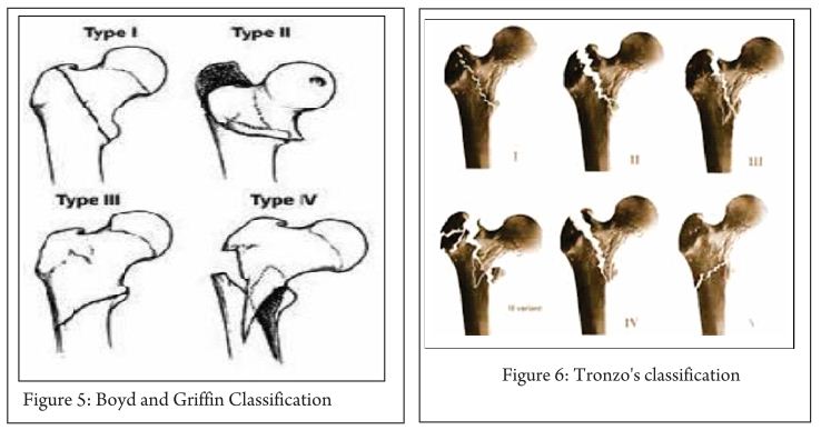
They were first to mention instability in both coronal and sagittal plane. This classification, included fractures from the extracapsular part of the neck to a point 5 cm distal to the lesser trochanter.
Type 1: Fractures that extend along the intertrochanteric line.
Type 2: Comminuted fractures with the main fracture line along the intertrochanteric line but with multiple secondary fracture lines (may be in coronal plane).
Type 3: Fractures that extend to or are distal to the lesser trochanter.
Type 4: Fractures of the trochanteric region and proximal shaft with fractures in at least two planes.

Clinical importance:
Type 1- Reduction usually is simple and is maintained with little difficulty. Results generally are satisfactory
Type 2- Reduction of these fractures is more difficult because the comminution can vary from slight to extreme
Type 3- these fractures usually are more difficult to reduce and result in more complications at operation and during convalescence.
Type 4- if open reduction and internal fixation are used, two-plane fixation is required because of the spiral, oblique, or butterfly fracture of the shaft.

Tronzo’s classification [7] (1973) (Fig. 6):

Tronzo incorporated Boyds and Griffin two plane instability in classification.
Type 1: Incomplete fractures
Type 2: Uncomminuted fractures, with or without displacement; both trochanters fractured
Type 3: Comminuted fractures, large lesser trochanter fragment; posterior wall exploded; neck beak impacted in shaft
Type 3 Variant: As above, plus greater trochanter fractured off and separated
Type 4: Posterior wall exploded, neck spike displaced outside shaft
Type 5: reverse obliquity fracture, with or without greater trochanter separation

Clinical importance:
This system is complex to use & not adequate to apply in clinical practice. It has poor reliability, though can be used for documentation of long-term results and comparison of treatment modality. Yet many surgeons prefer it for its simplicity and biomechanical rationale.

The Ramadier’s Classification[8](Fig. 7):Fig 7
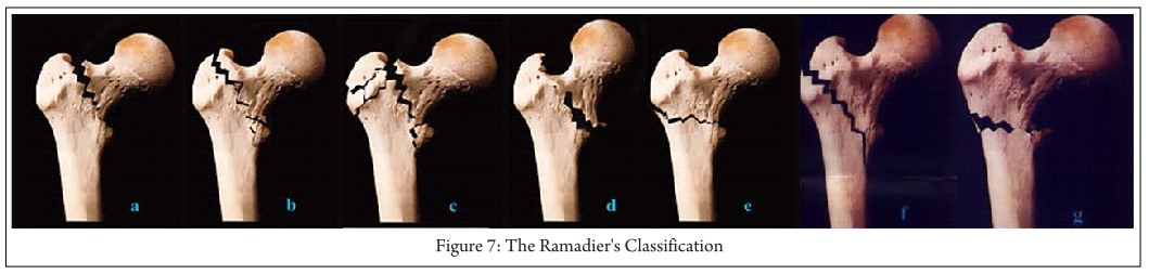
A: Cervico-trochanteric fractures- with a fracture line at the base of the femoral neck
b: Simple pertrochanteric fractures- fracture line that runs parallel to the intertrochanteric line; frequently, the lesser trochanter is broken off
c: Complex pertrochanteric fractures have an additional fracture line that separates most of the greater trochanter from the femoral shaft; the lesser trochanter is often fractured
d: Pertrochanteric fractures with valgus displacement- fracture line that begins on the greater trochanter and finishes below the lesser trochante
e: Pertrochanteric fractures with an intertrochanteric fracture line
f: Trochantero-diaphyseal fractures- spiral line through the greater trochanter and into the proximal shaft often with 3rd fragment.
G: Subtrochanteric fractures- more or less horizontal fracture line that runs below the two trochanters

Decoulx and Lavarde’s classification [9](1969):Simple anatomical classification for descriptive purposes.
Cervico-trochanteric fractures
Pertrochanteric fractures
Intertrochanteric fractures
Subtrochanteric fractures
Subtrochantero-diaphyseal fractures
The Briot Classification Diaphyseo-Trochanteric Fractures [10] (1980) (Fig. 8):Fig 8 9
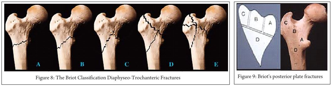
A Evans’ reversed obliquity fracture
B “Basque roof” fractures
C Boyd’s “steeple” fracture
D Fractures with an additional fracture line ascending to the intertrochanteric line
E Fractures with additional fracture lines radiating through the greater trochanter
Briot’s posterior plate fractures
Note: Boundaries of posterior plate, Maximum extent of plate, Possible fracture lines
Clinical importance: Its simple and based on biomechanical concept. Briot’s found posterior wall fracture is important for sagittal instability and external rotation sometimes causing malunion in external rotation. Reduction can be done in these by internal rotation reducing the anterior gap while realign the posterior fractured wall.

Ender Classification(1970)[11] (Fig. 10):Fig 10
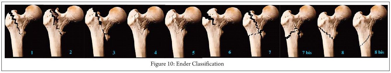
Trochanteric eversion fractures
-1. Simple fractures
-2. Fractures with a posterior fragment
-3 Fractures with lateral and proximal displacement
3. trochanteric inversion fractures
-4. With a pointed proximal fragment spike
-5 .With a rounded proximal fragment beak
6. Intertrochanteric fractures
Subtrochanteric fractures
-7 and 7a Transverse or reversed obliquity fractures
-8 and 8a Spiral fractures
Clinical importance: This classification gives information on injury mechanism, which can be helpful to reduce fracture while performing closed nailing.

Dr G. S. Kulkarni et al Classification / Modified Jenson-Evan’s Classification[1] (Fig 11): Fig 11
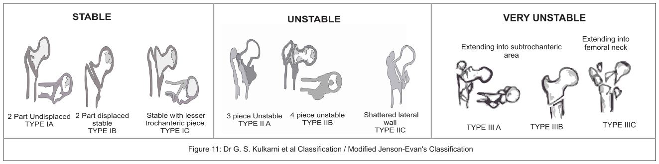
Dr G.S. Kulkarni et al [1] published his new classification in intertrochanteric fractures based on AO & Evan-Jansen classification. He added new varieties of intertrochanteric fractures described by Gotfried[12] and Kyle [4]. This classification is treatment oriented and will help in deciding the implant according to the fracture type.
Type IA- stable undisplaced.
Type IB- stable minimally displaced.
Type IC- stable minimally displaced with a small fragment of lesser trochanter.
Type IIA- unstable 3 piece fracture with large posteromedial fragment of lesser trochanter.
Type IIB- 4 piece fracture.
Type C- Shattered lateral wall.
Type IIIA- trochanteric fracture with extension into subtrochanter.
Type IIIB- reserve oblique.
Type IIIC- trochanteric fracture with extension into femoral neck area.

Clinical Importance: Classification helps in selecting treatment protocols as below.
Type I: This stable fractures can be managed by any fixation modality gives excellent results. DHS is implant of choice.
Type II: These unstable fractures are described as problem fractures can be managed with DHS with some modification or IMN.
Type III: This very unstable fracture with DHS gives poor results. In these type with lateral wall fracture use of DHS lead to excessive collapse, pain, restricted mobility in hip, sometime non union and failure. Intramedullary nails (IMN) are better choice as they prevents excessive collapse at fracture site, better restoration of anatomy and biomechanically stronger implants; Arthroplasty can also be done in select cases. Unusual fracture pattern like basi-cervical fractures extension can be fixed with additional derotation screw as these are also rotationally unstable. Reverse oblique pattern like fracture lateral wall are better fixed with IMN.
Conclusion:
Various classifications have been proposed over years described the fracture patterns, focusing on importance of posteriomedial and lateral wall for stability. Tronzo classification is found to be less reliable and not useful in clinical practice. AO/OTA and Dr G.S. Kulkarni et al modified classification has described in detail the preferred implant according to the fracture type. An AO/OTA group has good reliability but subgroup assessment has poor reliability; it is more useful in record keeping, deciding management and research. Kulkarni et al classification is found to be more simple & easy to apply in practice, record keeping and research. There is still no consensus on the best classification but with new biomechanical informations coming through, the classification systems would continue to evolve.


References

1. GS Kulkarni, Rajiv Limaye, Milind Kulkarni, Sunil Kulkarni. Current Concept review: Intertrochanteric fractures. Indian Journal of Orthopaedics.2006;40:16-23.
2. Evans, E. M. () The treatment of trochanteric fractures of the femur. J. Bone Jt Surg. 1949;31-B: 190-203.
3. Jensen J. S. Classification of trochanteric fractures. Actaorthop. Scand. 1980; 51:803-810.
4. Kyle R. F., Gustilo R. B. And Premer R. F. Analysis of six hundred and twenty-two intertrochantenc hip fractures. J. Bone joint(Am). 1979;61: 216-21.
5. M.E. Muller, S. Nazarian, P. Koch, J. Schatzker The comprehensive classification of fractures of long bones Springer, Berlin. 1990.
6. Boyd HB, Griffin LL. Classification and treatment of trochanteric fractures. Arch Surg. 1949; 58:853.
7. Tronzo RG. Symposium on fractures of the hip. Special considerations in management. Orthop Clin North Am. 1974; 5(3): 571–583.
8. M. Bombart, J.O. Ramadier Trochanteric fractures Rev Chir Orthop, 52 (1966), 353–374.
9. Decoulx P, Lavarde G. Fractures of the trochanteric region. A statistical study of 2,612 cases. J Chir (Paris). 1969; 98(1):75-100.
10. Briot B. Fractures per-trochantériennes: anatomie pathologique et classification. Cahiers d’Enseignement de la SOFCOT Expansions Sci Franc.1980;12: 69-76.
11. J. Ender Per- und subtrochantere Oberschenkelbrüche. Hefte Unfallheilk;1970:106, 2–11.
12. Gotfried Y. The lateral trochanteric wall. Clin Orthop. 2004; 425:.82-86.


How to Cite this article: Sonawane DV. Classifications of Intertrochanteric fractures and their Clinical Importance. Trauma International July-Sep 2015;1(1):7-11

Authors


(Abstract)      (Full Text HTML)      (Download PDF)


Intramedullary Nail Versus Dynamic Hip Screw; Intramedullary Nail (Advantages And Disadvantages)

Vol 1 | Issue 1 | July – Sep 2015 | page: 17-20 | Ajay Pal Singh[1], Vivek Kochar[2]


Author: Ajay Pal Singh[1], Vivek Kochar[2].

[1] Punjab Civil Medical Services-1, Civil Hospital Mukerian, Punjab, India.
[2] Healing Touch hospital, Ambala, Haryana. India..

Address of Correspondence
Dr. Ajay Pal Singh,
Kanwar Hospital, Hoshiarpur, Punjab, India.
Email: docajaypal@gmail.com


Abstract

since 1950s surgical treatment of extracapsular hip fractures is done  using a variety of different implants. Unstable fractures  are fractures with lateral wall or posteromedial comminution,fractures with reverse obliquity patterns and fractures extending into the femoral neck or subtrochanteric regions. These types are not well controlled by the sliding compression hip screw and side plate and  are associated with a high rate of fixation failure when treated with dynamic hip screw. Theoretical biomechanical advantages of  intramedullary nails over screw and plate fixation are attributed to a reduced distance between the hip joint and the implant. Success of Proximal femoral nail for the treatment of such fractures is based on biomechanical principles,cadaver studies and clinical series. Although it is seems that  nail fixation is superior to sliding compression hip screw and side plate fixation for the treatment of unstable fractures, this point is not well proven till date. Orthopedic literature does not support the superiority of intramedullary nail fixation over  sliding hip screw fixation for the treatment of intertrochanteric femoral fractures and intramedullary nail fixation is associated with a higher complication rate. Till date the debate over superiority of extramedullary fixation versus intramedullary implants continues especially in unstable intertrochanteric fractures. This article highlights the advantages and disadvantages of proximal femoral nail in intertrochanteric fractures with review of literature.
Keywords: Intertrochanteric fractures, intramedullary nail, dynamic hip screw


Introduction

Intertrochanteric hip fractures account for approximately half of the hip fractures in the elderly and pose a number of management dilemmas depending on the fracture configuration and status of the bones. A wide variety of implants are available for the internal fixation of these fractures ranging from dynamic hip screw which can be combined with trochanteric stabilisation plate; locking plates; intramedullary implants such as proximal femoral nail (PFN), Gamma nail.
In the last century dynamic hip screw used to be the gold standard treatment for intertrochanteric fractures. But in the last 2 decades more surgeons now prefer proximal femoral nails for these fractures. There has been a 20 fold increase in the use of intramedullary nails in America since 1999 [1]. However, Cochrane review of 2010
found dynamic hip screw (DHS) to be superior to intramedullary nails in all trochanteric fractures [2]. So why this change? Why more and more orthopaedicians are shifting to fixation of these fractures with an intramedullary nail?

Background
A search of the literature for the ideal implant in intertrochanteric fractures in the review groups of Cochrane library favors the use of sliding hip screw over intramedullary implants [2,3,4]. Also reviews of various other meta-analysis were in favor of sliding hip screw [1,5,6]. This was predominantly due to risk of femoral shaft fracture associated with earlier version of gamma nail and complications due to steep learning curve associated with the implant [7, 8]. The aim in the treatment of intertrochanteric fractures is not only to achieve fracture union but restoration of mobility and function in the shortest duration of time with minimal complications. Fig. 1 is a radiograph of fixation of an unstable fracture with dynamic hip screw and shows break of the lateral femoral wall intraoperatively resulting in medialization of femoral shaft with shortening with high chances of screw cut out with mobilization. In all these patients our primary aim of early mobilization is defeated and we end up keeping the patient in bed for a longer time.Fig 1
Re-operation rates of 4–12% have been reported following the gold standard technique of fixation with dynamic hip screw [9-11]. The re-operation rates are particularly high in patients with unstable fractures. Re-operations are usually performed for medialization of the femoral shaft following mobilization of the patients [9-12].
So considering such high rates of complications in unstable intertrochanteric fractures and the biomechanical advantages of intramedullary implants has made PFN an attractive option. With improved surgical techniques and improved designs of the nail, studies have shown promise in terms of decrease in the rate of complications and early return to pretrauma mobility status [13,14].

Advantages of Intramedullary nails
Less chances of screw cut out
Load bearing in the proximal femur is predominantly through calcar femorale, the lever arm of laterally placed plate is increased so there is a risk of implant cut out. [15](Fig 2).
Biomechanically, compared to a laterally fixed side plate, an intramedullary device decreases the bending force of the hip joint on implant by 25-30%. This has advantages especially in elderly patients, in whom the primary treatment goal is immediate full-weight bearing mobilization.
Implant of choice in lateral wall compromised fractures
Traditionally it was the posteromedial comminution which was considered the most important factor in determining the severity of fracture. The importance of the integrity of the lateral femoral wall has been documented recently [9,10,12]. The lateral wall is the proximal extension of the femoral shaft. This lateral wall is extremely thin in unstable 31-A2 type fracture [9,10]. The lateral wall in patients treated with dynamic hip screw provides a lateral buttress for the controlled fracture impaction and preventing collapse. Palm et al found that there was eight times higher risk of reoperation due to technical failure with the gold standard technique of dynamic hip screw in patients with fracture of the lateral femoral wall [9] This has been attributed to the fact that when the lateral femoral wall is fractured, the fracture line is parallel to the sliding vector of the sliding hip screw, which, as in the reverse oblique intertrochanteric fracture, allows the trochanteric and femoral head and neck fragments to slide laterally and the shaft to slide medially. The fracture complex subsequently disintegrates, with a high risk of failure including cutout of the screw into the hip joint.Fig 2
Another fact is that most of the fractures of the lateral femoral wall occurs intraoperatively with the gold standard technique when the large diameter hole is drilled into the lateral femoral wall, thereby converting a 31-A2 type to 31-A3 type. Gotfried in a retrospective analysis of twenty-four patients with documented postoperative fracture collapse and there findings showed unequivocally that in all patients, this complication followed fracture of the lateral wall and resulted in protracted period of disability until fracture healing [10] The importance of the integrity of the lateral wall for event-free fracture healing clearly is indicated, and fracture of the lateral wall should be avoided in any fixation procedure.
Palm et al have recommended dividing the fractures into two categories: A1 to A2.1 & A2.2 to A3`, and not just into A1, A2, and A3 fracture types as has been reported in most studies taking into account the integrity of lateral femoral wall [9]. This has implication on treatment guideline that the dynamic hip screw is not a good implant in patients falling into the second category. In the series by Gotfried, lateral wall fracture occurred in a third of the hips with the most vulnerable lateral femoral wall i.e., in those with an AO/OTA A2.2 or A2.3 fracture, which lacks buttress support of the greater trochanter [10]
In these fractures with compromised lateral wall, either a locking plate (proximal femoral locking compression plate or reverse distal femoral locking compression plate) or DHS with trochanteric stabilization plate which acts as lateral buttress and limits excessive collapse. The other option is an intramedullary nail which acts by bypassing the lateral wall and acts as a prosthetic lateral cortex medial to the broken lateral wall. Most of the recent studies suggest PFN as gold standard implant in these type of fractures [16,-18](Fig.3)
Less Blood loss during Surgery/ Less operative time/ Smaller incision
Zhang et al in an extensive meta-analysis of 6 randomised and quasirandomised studies concluded that PFN group had significantly less operative time, intraoperative blood loss), and length of incision than the DHS group [14].

Limb length shortening
Intramedullary nails are associated with less shortening and less sliding of the lag screw. This is due to the fact that intramedullary nail stops the telescoping displacement of the proximal aspect of the femur [19].  In fact, the proximal part of the nail blocks the head-and-neck fragment, preventing its complete impaction.

Disadvantages of intramedullary nails

Implant failure
Implant failure after fixation of intertrochanteric fractures has been reported to be quite high in the tune of approximately 20% in various series [20-22]. Implant failure can be in the form of broken nail (Fig. 4), Z effect with medialization of superior screw and lateralization of inferior screw on weight bearing in earlier versions of PFN with 2 proximal screws, Reverse Z effect in which superior screw goes laterally and inferior screw goes medially (Fig. 5) and screw cut out with varus collapse (Fig.6a, 6b).
This can be attributed to steep learning curve with intramedullary nail fixation of these fractures. Secondly most of the series have been reported in unstable intertrochanteric fractures thus leading to bias. But with improvement in the surgical techniques and improved design of the nail trying to be as similar to the geometry as possible, results have improved [13, 14].  But the most common reason for implant failure is inability to achieve proper reduction.
Recent meta analysis by the Cochrane comparing different designs such as screw nail combinations to helical blade nail combinations have shown no significant differences [23].  But in our experience the results have improved significantly in our hands with the Proximal Femoral Nail Antirotation II. But it still requires more evidence to recommend helical blade usage for routine use.

Non Union
Nonunion without implant failure is a rare complication (Fig.7). And the most common reason is improper reduction. Intertrochanteric fractures with posterior sag or fractures with coronal split are the most likely to go for non union. Another reason is fixation of fracture with distraction at the fracture site.

Shaft femur fracture at nail tip
Generally short PFN are straight nail which when used in osteoporotic bones may impinge on the anterior femoral cortex which may result in fracture due to excessive force used while inserting the nail (Fig. 8a, 8b). It is a very rare complication but one has to be very careful to avoid this complication.

High cost of implant
Cost of an intramedullary nail is 7-8 times the cost of a dynamic hip screw which may act as a restraint for use in all patients with intertrochanteric fractures. So there is still a place for DHS in intertrochanteric fractures especially stable ones. But in unstable fractures, DHS needs to be supplemented with trochanteric stabilization plate or we need a PFLCP or reverse DFLCP which costs more to the patient. So cost of implant should not be a factor in unstable intertrochanteric fractures.

Conclusion
Advantages of proximal femoral nail are less surgical trauma, less screening time,
less blood loss and earlier rehabilitation, the ease of implantation and the possibility of early weight-bearing even after very complex fractures. Surgical expertise is necessary to avoid the complications associated with PFN.


References

1. Anglen JO, Weinstein JN. Nail or plate fixation of intertrochanteric hip fractures: changing pattern of practice. A review of the American Board of Orthopaedic Surgery Database. J Bone Joint Surg Am. 2008;90:700–7.
2. Parker MJ, Handoll HH. Gamma and other cephalocondylic intramedullary nails versus extramedullary implants for extracapsular hip fractures in adults. Cochrane database Syst Rev [Internet]. 2010 Jan [cited 2015 Mar 8];(9):CD000093.
3. Parker MJ, Handoll HHG. Gamma and other cephalocondylic intramedullary nails versus extramedullary implants for extracapsular hip fractures in adults. Cochrane database Syst Rev [Internet]. 2008 Jan [cited 2015 Mar 8];(3):CD000093.
4. Parker MJ, Handoll HHG. Gamma and other cephalocondylic intramedullary nails versus extramedullary implants for extracapsular hip fractures. Cochrane database Syst Rev [Internet]. 2004 Jan [cited 2015 Mar 8];(1):CD000093.
5. Jones HW, Johnston P, Parker M. Are short femoral nails superior to the sliding hip screw? A meta-analysis of 24 studies involving 3,279 fractures. Int Orthop. 2006;30:69–78.
6. Jiang S-D, Jiang L-S, Zhao C-Q, Dai L-Y. No advantages of Gamma nail over sliding hip screw in the management of peritrochanteric hip fractures: a meta-analysis of randomized controlled trials. Disabil Rehabil. 2008;30:493–7.
7. Radford PJ, Needoff M, Webb JK. A prospective randomised comparison of the dynamic hip screw and the gamma locking nail. J Bone Jt Surgery, Br Vol [Internet]. 1993;75-B:789–93.
8. O’Brien PJ, Meek RN, Blachut PA, Broekhuyse HM, Sabharwal S. Fixation of intertrochanteric hip fractures: Gamma nail versus dynamic hip screw. A randomized, prospective study. Can J Surg. 1995;38:516–20.
9. Palm H, Jacobsen S, Sonne-Holm S, Gebuhr P. Integrity of the lateral femoral wall in intertrochanteric hip fractures: an important predictor of a reoperation. J Bone Joint Surg Am. 2007;89:470–5.
10. Gotfried Y. The lateral trochanteric wall: a key element in the reconstruction of unstable pertrochanteric hip fractures. Clin Orthop Relat Res. 2004;82–6.
11. Haidukewych GJ, Israel TA, Berry DJ. Reverse obliquity fractures of the intertrochanteric region of the femur. J Bone Joint Surg Am. 2001;83-A:643–50.
12. Im G-I, Shin Y-W, Song Y-J. Potentially unstable intertrochanteric fractures. J Orthop Trauma. 2005;19:5–9.
13. Pu JS, Liu L, Wang GL, Fang Y, Yang TF. Results of the proximal femoral nail anti-rotation (PFNA) in elderly Chinese patients. Int Orthop. 2009;33:1441–4.
14. Zhang Kairui, Zhang Sheng, Yang Jun, Dong Weiqiang, Wang Shengnan CY, Al-Qwbani Mohammed, Wang Qiang Y Bin. Proximal Femoral Nail vs.Dynamic Hip Screw in Treatment of Intertrochanteric Fractures: A Meta-Analysis. Med Sci Monit. 2014;20:1628–33.
15. Banan H, Al-Sabti A, Jimulia T, Hart AJ. The treatment of unstable, extracapsular hip fractures with the AO/ASIF proximal femoral nail (PFN)–our first 60 cases. Injury [Internet]. 2002 Jun [cited 2015 Mar 8];33(5):401–5.
16. Yao C, Zhang CQ, Jin DX, Chen YF. Early results of reverse less invasive stabilization system plating in treating elderly intertrochanteric fractures: A prospective study compared to proximal femoral nail. Chin Med J (Engl). 2011;124:2150–7.
17. Zhou F, Zhang ZS, Yang H, Tian Y, Ji HQ, Guo Y, et al. Less Invasive Stabilization System (LISS) Versus Proximal Femoral Nail Anti-rotation (PFNA) in Treating Proximal Femoral Fractures. Journal of Orthopaedic Trauma. 2012. p. 155–62.
18. Haq RU, Manhas V, Pankaj A, Srivastava A, Dhammi IK, Jain AK. Proximal femoral nails compared with reverse distal femoral locking plates in intertrochanteric fractures with a compromised lateral wall; a randomised controlled trial. Int Orthop [Internet]. 2014 Jul [cited 2015 Mar 8];38(7):1443–9.
19. Hardy DC, Descamps PY, Krallis P, Fabeck L, Smets P, Bertens CL, Delince PE. Use of an intramedullary hip-screw compared with a compression hip-screw with a plate for intertrochanteric femoral fractures. A prospective, randomized study of one hundred patients. J Bone Joint Surg Am. 1998 May;80(5):618-30.
20. Sadowski C, Lübbeke A, Saudan M, Riand N, Stern R, Hoffmeyer P. Treatment of reverse oblique and transverse intertrochanteric fractures with use of an intramedullary nail or a 95 degrees screw-plate: a prospective, randomized study. J Bone Joint Surg Am. 2002 Mar;84-A(3):372-81.
21. Gavaskar AS, Subramanian M, Tummala NC. Results of proximal femur nail antirotation for low velocity trochanteric fractures in elderly. Indian J Orthop. 2012 Sep;46(5):556-60.
22. Saudan M, Lübbeke A, Sadowski C, Riand N, Stern R, Hoffmeyer P. Pertrochanteric fractures: is there an advantage to an intramedullary nail?: a randomized, prospective study of 206 patients comparing the dynamic hip screw and proximal femoral nail. J Orthop Trauma. 2002 Jul;16(6):386-93.
23. Queally JM, Harris E, Handoll HHG, Parker MJ. Intramedullary nails for extracapsular hip fractures in adults. Cochrane database Syst Rev [Internet]. 2014 Jan [cited 2015 Mar 8];9:CD004961.


How to Cite this article: Singh A P, Kochar V. Intramedullary Nail Versus Dynamic Hip Screw; Intramedullary Nail (Advantages And Disadvantages). Trauma International July-Sep 2015;1(1):17-20

Author


(Abstract)      (Full Text HTML)      (Download PDF)


Complications Related to Intertrochanteric fractures

Vol 1 | Issue 1 | July – Sep 2015 | page: 25-30 | Vaibhav Bagaria[1], Farokh Wadia[2]


Author: Vaibhav Bagaria[1], Farokh Wadia[2].

[1] Department of Orthopedics, Sir HN Reliance Foundation Hospital,
Mumbai, India.

Address of Correspondence
Dr Vaibhav Bagaria
Department of Orthopedics, Sir HN Reliance Foundation Hospital, Mumbai, India.
Email: bagariavaibhav@gmail.com


Abstract

Intrertrochanteric fractures are not very prone for complications, however when complications do occur they are quite disabling and challenging. Major complications can be avoided by understanding the personality of the fracture and by choosing correct implant. Complications can be listed as implant cut out, non union, implant failure, peri implant fracture and general medical complications. In this review we present the current evidence related to complications of Intertrochanteric fracture and also certain guidelines to avoid these complications
Keywords: Intertrochanteric fractures, complications, failure


Introduction

Inter trochanteric fractures and their complications are some of the commonest conditions seen by an orthopedic surgeon in their clinical practice. While some of these complications are avoidable and by optimization of technique and resources the outcomes can be improved, there are others which are unavoidable in a given circumstances. Since these fractures typically occur in geriatric group and the patient is often frail and having comorbidities, complications in this groups leads to loss of functional independence, prolonged hospital stay with tremendous costs to the patients, family and the society. The outcome after Inter trochanteric fracture especially in the elderly is dependent on several factors and the best way to manage complications is to preempt them. The paradigm is shifting for intertrochanteric fractures globally. The older paradigm, in the 20th century, of quick surgery and just getting the fracture to heal is no longer acceptable for most patients who are expected to have an independent ambulatory capacity and dignified quality of life

A thorough understanding of the possible complications and why they happen is cornerstone of attempts in reducing them. This chapter focuses on how these complications can be classified, evaluated, prevented and managed in cases where in these happen. There are several ways in which these complications can be classified. One of the ways is to associate them with their cause and by doing this, one can help prevent them and formulate a strategy where in the cause and management is protocolized. The complications can be Fracture related; Related to choice of the implant; Routine operative and general complications.

Materials and Methods:
AFracture pattern Related:
Stable Inter trochanteric fractures do well with any kind of fixation. On the other hand the unstable patterns. Complication rates such as screw cut outs have been reported to be as high as 50% in cases of unstable IT fractures ( 1) There have been some recent insights with regard to fracture pattern which have drastically improved our understanding of intertrochanteric fractures. The following concepts are discussed:
Fracture classification
Lateral wall fractures
Rotational instability
Fracture reduction

Fracture classification:
Previously used classification system of Boyd & Griffiths had poor inter and intra-observer reliability and were used commonly to describe fracture patterns rather than predict outcomes. Evans classification subsequently advanced our understanding about stable and unstable fracture patterns. But the most commonly used classification currently is the AO/OTA classification, which classifies IT fractures into three types: 31A1, 31 A2 and 31A3 with increasing instability as the grade increases. However, more important would be to classify them into two categories: 31A1.1 to 31A2.1 and 31A2.2 to 31 A3.3. The reason for this distinction as highlighted by Palm et al and Gotfried et al, is the quality of the greater trochanter and potential for a lateral wall fracture while inserting the sliding screw leading to a potentially unstable situation post-operatively.Fig 1

Lateral wall fractures:
Lateral wall is defined as part of the femoral cortex distal to the vastus ridge. Several studies have found that an inter-trochanteric fracture with extension into the lateral wall either existing or produced during insertion of screw plate device, leads to an unstable situation that cannot be salvaged with a sliding compression screw plate device. The latter are often labeled as iatrogenic lateral wall fractures (ILWF).
The support from lateral femoral wall is the key to success in sliding screw plate device to allow controlled compression and collapse at the fracture site. When the lateral femoral wall is fractured, the fracture line is parallel to the sliding vector of the sliding hip screw, which, as in the reverse oblique intertrochanteric fracture, allows the trochanteric and femoral head and neck fragments to slide laterally and the shaft to slide medially. The fracture complex subsequently disintegrates, with a high risk of failure. Palm et al in their series of 214 patients, found that a lateral wall fracture increases the risk of re-operation about 8 times, due to technical failure when a sliding hip screw was used. The bottom line is if there is a lateral wall or a greater trochanter fracture, sliding hip screw device should not be used.Fig 2

Rotational stability:
Until recent times, rotational stability of femoral head has been more or less neglected. With sliding compression screw devices, with single screw in the femoral head, the femoral head has been shown to be rotationally unstable especially with collapse and erosion of the neck with eventual cut out of the implant. To overcome this issue, many of the recent implants have added an extra screw incorporated within the plate design such as the PCP or the hybrid proximal femoral plate as well as the Inter TAN (Trochanteric antegrade nail) nailing system.

Fracture reduction:
Conventional teaching suggests that intertrochanteric fractures are reduced in internal rotation on fracture table. However, studies by Bannister et al and May et al show that up to 23% of intertrochanteric fractures reduce in external rotation. Excessive internal rotation can cause fracture gapping posteriorly which adds to destabilizing the fracture which already has postero-medial comminution.
Another common error is to accept less than optimal reduction. The most common malreduction is a posteriorly sagging femur shaft with an anterior step off at the fracture site. This may not be reduced by closed means and Carr et al [2] have described an open reduction maneuver where the shaft is first pulled laterally with a bone hook around the shaft to disimpact the fracture and then using a lever to push the head and neck fragment posteriorly to align with the shaft. This simple reduction maneuver is important to achieve anteromedial stability so as to allow immediate mobilization without putting excessive load on the implant.Fig 3

Implant Choice and complications: Three chief categories of implant used for these fractures are sliding/ dynamic hip screw, Intramedullary nails and endo prosthesis.
Dynamic Hip Screw & Medoff Plate: Cochrane review by Parker et al ( 3) have demonstrated the superiority of sliding hips screw in the management of the extra capsular neck fractures. In the review the studies included twenty-two trials (3749 participants) that compared the Gamma nail with the sliding hip screw (SHS). The Gamma nail was associated with increased risk of operative and later fracture of the femur and increased reoperation rate. There were no major differences between implants in wound infection, mortality or medical complications. Five trials (623 participants) compared the intramedullary hip screw (IMHS) with the SHS. Fracture fixation complications were more common in the IMHS group. Results for post-operative complications, mortality and functional outcomes were similar in both groups. Three trials (394 participants) showed no difference in fracture fixation complications, reoperation, wound infection and length of hospital stay for proximal femoral nail (PFN) versus the SHS.
Intramedullary femoral nails: There has been an increase in the use of these devices in last decades up from 3% to 67% as observed by Anglen et al in their study (4).
They have a distinct advantage over DHS when it comes to unstable inter trochanteric fractures. They however do have a complication set of their own which includes iatrogenic femoral fractures; difficult initial reduction and persistent thigh pain due to stress concentration around nail tip, varus angulation and secondary risk of femoral fractures. The classical indication for nailing is in managing the unstable patterns that include reverse obliquity fractures, transtrochanteric fractures, fractures with a large posteromedial fragment implying loss of the calcar buttress, and fractures with subtrochanteric extension. The reason for favoring nails in tis cases is based on biomechanical fact that since a nail is located closer to the centre of gravity and force transmission, the lever arm is shorter and there is less stress on the implant. They can thus resist higher forces across the medial calcar than what a sliding hip screw can. Intramedullary placement also prevents shaft medialization, which may commonly happen with unstable fracture patterns.

While there are a variety of devices that can be included in this category, the two commonest devices used are Gamma nail (Stryker) and Proximal Femoral Nail ( AO Synthes). The Gamma Nail was introduced before the PFN, which in comparison to a GN had a longer length, had two proximal screws and had a smaller diameter and could thus be inserted un reamed. The addition of an additional anti rotation screw was thought to decrease the probability of screw cut outs. The study by Schipper et al however did not find any difference in the cut out rates. It was then hypothesized that the so-called ‘knife effect’ counterbalances any advantage that the derotation screw offers in preventing the cutouts. The ‘ knife effect’ or the ‘Z effect’ originally described by Werner et al refers to development of condition following fixation with PFN in which the superior smaller screw migrates medially and the distal larger screw migrates laterally. It is believed that the medial cortex communition and a varus reduction is a contributory factor in this phenomenon (5).

Chief Complications with IM Devices:

1. Fractures of femoral Shaft:
2. Failure of fixation
3. Complication Associated with Distal Locking
The recent Cocharane database review 2014 that compare the various different types of intramedullary nails suggest that there is insufficient data to clearly state whether there are any outcome difference with the use of different types of intramedullary devices. It also states that since there is a clear evidence of superiority of the sliding hip screw over the intramedullary nail in the management of extra capsular fracture, further studies on different designs of these nails should take a back seat and a comparative trial for any new design should be made against the clearly superior sliding hip screw [6].

Primary Endo Prosthesis: A certain section of orthopedic surgeons believe that the primary arthroplasty for these procedures may help in early mobilization and consequently reduce mortality and morbidity associated with prolonged immobilization especially pulmonary atelectasis pressure sores and venous thromboembolism. While this rationale may not be applied to all cases, there is a small set of fracture patterns and patient groups in which the arthroplasty can play a role. In most cases they are usually the salvage procedure after secondary complications of conventional fixations. Hassankhani et al (7) in their study of 80 patients found hemiarthroplasty as a superior alternative with reduced rates of complications and patients in elderly patients who had unstable intertrochanteric fractures.
The key techniques that need to be remembered specifically for these cases are 1. Ensuring that the prosthesis is inserted or sunk to a point on stem that has been marked explicitly before insertion. It is important that the lesser trochanter as the land mark may not be an option in many of these cases. This helps in reducing dislocations and ensuring limb length symmetry.
2. Use of cement in cases of wide medullary canal and appropriate precautions during cementing to be taken.
3. Reconstruction of lesser trochanter with cables after the prosthesis is fixed.
4. Greater Trochanter reconstruction with use of K wires, cannulated screw or using tension band principle.

If done well, the studies by Sancheti et al a has revealed that results are comparable to fracture fixation and the only complication is of bed sore [8].

Major complications following the intertrochanteric fracture fixation surgery are:
A. Implant cut-out
This still remains the commonest cause of failure of the implant-fracture construct. It is an accepted almost dogmatic mandate that the tip apex distance correlates directly with implant cut-out, with higher the TAD, the greater is the risk of cut-out. This applies definitely for the sliding hip screw and has been shown recently to apply for cephalomedullary devices too.

Key technical points in preventing Implant Related complications:

1.Use of Tip Apex distance (TAD)& Parker ratio: Baumgaertner et al [9,10] first described the concept of TAD. This is a useful indicator for an optimal screw placement spatially irrespective of whether a plate or a nail is used. It is considered one of the most important indicators of hardware placement in most studies done on the failure of devices. Conventional thinking propagated a slight posterior and inferior placement of screw based on the philosophy that it gives more area for the screw to cut out. However the studies have shown that this increases the TAD and can be actually detrimental. Based on the concept it is agreed that a screw placed as central and as deep without perforating the head (within 10 mm of subchondral bone) is the ideal placement. Numerically speaking trauma surgeons should aim for the TAD of around 20 mm, generally the cases with TAD less than 25 mm have good outcomes.

Parker Ratio:
This is a mathematical measurement of screw placement within the femoral head and is a ratio of AB to AC multiplied by 100 in both AP and Lateral view. Parker in his original series of 225 patients with screw cutout in 25 found that on the AP view the average ratio was 45 for union and 58 for cut-out and it was 45 for union and 36 for cut-out on the lateral view.

Tip apex distance may not be valid for all types of implants. The proximal femoral nail antirotation (PFNA) is unique with a helical blade proximally that is impacted in the cancellous bone of the femoral head instead of using a standard lag screw. While this allows a better purchase in the femoral head and provides rotational stability, they have also been associated with increased cut out albeit for a different reason. Nikoloski et al [11]found that the tip apex distance of less than 20 mm for these devices was found to be associated with a higher incidence of blade cut out due to penetration of the subchondral bone of the femoral head
2. Assessment of the lateral wall: If there is a lateral wall fracture then the choice of implants becomes crucial. These fractures could either be reverse obliquity inter trochanteric fractures or trochanteric fractures. In these fracture the medialization of the femoral shaft with a lateral migration of proximal fragment occurs. If these occur there is a high chance of mal reduction, non union and screw cut out. Intramedullary devices in these cases are most suitable. Sliding hip screw when used must be either a ‘medoff plate’ or in conjunction with trochanteric stabilization plate or in some cases locking plates.
3.Entry Point: A good entry point for trochanteric nails is slightly medial to the tip of the greater trochanter. This helps prevents varus angulation. It is also important that while reaming the proximal part the reamers are well inside the proximal part past the entry point so as not to enlarge the entry point laterally.
4. Respecting the femoral Bow: One of the commonest complications of intramedullary nail is the iatrogenic fracture created during the nail insertion. It is important to be aware of the femoral bow, to check fluroscopically if the nail is hitting against the anterior cortex and not to hammer the nail especially in the osteoporotic patients.
5. Avoiding Varus Angulation: Any varus angulation increase the lever arm, increasing the stress on the implant predisposing it to failure. A good way to judge this intra operatively is to look at relative positions of tip of the GT and the centre of femoral head. For a good alignment they should be co planar. If the GT tip is higher than the centre of femoral head, it is a varus reduction and vice versa if the tip is higher. In most cases use of 130 degree nail and sliding screw is appropriate but can vary from person to person.
6. Avoiding Fracture distraction: This is especially true with the use of nails in which it is not uncommon to see malrotation and distraction. If the device is fixed in a distracted position, there is insufficient osseous contact and some of the load that would have been normally borne by this contact is entirely borne by the device and places it in vulnerable position prone to fatigue failures. The nail tends to break at its weakest point, which is the largest apertures for the screw in the nail. To avoid distraction, it is important to release traction before locking and also confirming the same on image intensifier.

B. Non Union:
Non-union is very rare after inter-trochanteric fractures and has been reported in 1% of older population. The reasons for non-union are poorly understood as the fracture affects metaphyseal bone with huge cancellous surfaces. The speculated reasons include delayed treatment, unfavorable fracture pattern, poor bone quality, and suboptimal internal fixation. While salvage total hip replacement is the treatment of choice for elderly patients with non-union, the technique is more difficult and often special design implant such as calcar replacement prosthesis, extended neck stem or long stem implant is required. Haidukewych et al [12] described good results in a series of 60 patients with failed intertrochanteric fractures that underwent revision to total hip replacement or bipolar hemiarthroplasty. A total of five reoperations were performed: two patients had a revision, one had a rewiring procedure because of trochanteric avulsion, one had late removal of trochanteric hardware, and one had debridement of fat necrosis. One patient had two dislocations, both of which were treated with closed reduction. They reported a survival rate of 100% at 5 yrs and 89% at 10 years.
In younger patients, revision fracture fixation should be attempted. Dhammi et al [13] have reported a 100% union rate in 18 patients at a mean follow-up of 5.6 months with excision of pseudarthrosis, freshening of bone edges, stable fixation with a 135 degree DHS, valgization and bone grafting. Similarly Vidyadhara et al reported lateral closing wedge osteotomy with DHS fixation that resulted in union, with improvement in Harris hip scores from 34 to 89 in seven patients.

C. Implant Failure
Breakage of cephalomedullary implant is a rare but serious complication and has been reported to have an incidence of 2.9% in a series of 453 patients by Von Ruden et al. [14]In this series, the breakage occurred on an average of 6 months post-operatively. In majority, the cause was delayed or non-union due to insufficient initial reduction of the fracture.

D. Complication Associated with Cement Augmentation: While in the traditional method of fixations the failure in intertrochanteric fractures was commonly secondary to the screw cut out, in the cement-augmented group, this is relatively rare. In a study by Wu et al [15] of the 321 patients who under went Cemented augmented DHS fixation for their inter trochanteric fractures, no patient has a lag screw cut out. Six patients had delayed or non union with side plate failures, with three screw breakages, one plate breakage and one patient had infection and avascular necrosis each. The procedure related complications in this study was around 8.9%

E. Peri-implant fracture

Dorr has classified proximal femur based on morphology into three types:
Type A femur has a small metaphysis, thick cortex and high narrowed isthmus.
Type B femur has a wider metaphysis, thinner cortex and a tapering but wider isthmus
Type C femur has wide metaphysis, thin cortex and straight/varus curvature of diaphysis with wide isthmus.

There is a significantly higher risk of peri-prosthetic fracture in Type C femurs, especially intra-operative perforation with the use of long cephalomedullary device in a varus curvature femur.
Peri-prosthetic fractures are otherwise common around the distal interlocking screw of a cephalomedullary device. Marmor et al [16] in a biomechanical study of eighteen synthetic femora compared stiffness and load to failure of three nail lengths (short, extended short and long) and found that the axial stiffness was significantly higher for short nails compared to long nail design whereas extended short nail exhibited a significantly higher failure load than short nail constructs. All peri-prosthetic fractures occurred around the distal interlocking screw irrespective of nail length.

Intra Operative Complications:
Breakage of the guide wire
Perforation of the femoral head
Convergence of the guide wires
Intraoperative conversion to other method of fixation secondary to propagation of the fracture line,
F. General Operative & Medical Complications: Since majority of these fractures occur in elderly patients who have co morbidities and age related issue, the medical complications in pre and peri operative period is a major concern. It has been reported that there is 20% mortality in perioperative period in the patient who have had unstable fractures.

Venous Thrombo Embolism (VTE): Venous thrombo embolism which includes both deep vein thrombosis and pulmonary embolism is a relatively common occurrence after hip fracture surgery (17). There are various rates of incidence with significant variations depending upon the co morbidities, pre operative risk factors, and even the racial composition. In view of the preventable nature of the disease, routine pharmacologic thromboprophylaxis is recommended by many consensus groups. Although there is still controversy about the role of universal thrombo prophylaxis, there is large body of evidence that unequivocally suggests that the thrombo prophylaxis should be given to patients who were immobile for greater than 72 hours before the surgery, those who had obesity, history of malignancy and in those whom the surgery lasted more than 72 hours (18).

Blood Loss: Since majority of these fracture occur in the geriatric age group, it is very important that the blood loss be monitored and appropriately replinshed after the surgery. In many cases a pre operative transfusion to build up the hemoglobin is also required. Various studies have tried to quantify the amount of surgical blood loss that occur with various techniques. In one of the larger studies by Zhang et al [19] in which they calculated the obvious and hidden blood loss in 216 cases of inter trochanteric fracture treated by PFN and 168 cases treated by DHS, they found that PFN group had a mean loss of 48.9 +/- 2.8 ml during the procedure, an obvious blood loss of 62.3 +/- 3.8 ml and a hidden blood loss of 385 +/- 6.2 ml. In contrast the blood loss intra operatively during DHS fixation was 124.9 +/- 7.8 ml, the obvious blood loss was 73.9 +/- 4.7 ml and the hidden blood loss was 243.4 +/- 6.3 ml.

Osteoporosis associated complications: One of the commonest cause of both the intertrochanteric fracture and also its complication post surgical fixation is the presence of osteoporosis. In a study done by Bonnaire et al (20) the incidence of the screw cut outs have been linked directly to the presence of osteoporosis and decreased bone density at the bone trabeculae.

Compliance: It is very important that appropriate post surgery policy is formulated with regards to rehabilitation, weight bearing and regaining the bone strength.

Associated fractures: While treating intertrochanteric fractures it is also important to rule out any associated injuries. They may be associated with distal radius fracture and vertebral body collapses. Apart from a thorough initial assessment it is also important to take adequate precaution while positioning the patients.

Other Co morbidities: As mentioned previously many Neurovascular Damage: While the incidence of many major neuro vascular problem following the interrochanteric fracture fiation is rare, yet there may be complications related to in appropriate padding and issues with incorrect traction. There have been case reports of profunda femoris artery damage that needed vascular intervention ( Ref)

Wound Infections – Superficial and Deep: As is with any surgery there can be wound infection. This may be related to patient factors and general immunity of the patient, factors related to operating environment and also the duration of the surgery. It is important to distinguish between a superficial and deep infection. While local wound dressing and antibiotics may treat the superficial infection, a deeper infection may require repeated washouts, antibiotic beads and occasionally implant removal.

Hematoma formation: Many of the patients who suffer from this type of fracture are on anti coagulants. In view of the high incidence of the VTE in these surgery, many of these patients are also thromboprophylaxysed, increasing the chances of hematoma formation post operatively. It is thus important to ensure that a meticulous hemostasis is obtained intra operatively and local wound inspection is diligently maintained to pick up any early signs of development of a local hematoma.

Other general complications:
Pneumonia
Anesthesia Related complications:
Bed Sores
Cardiovascular
Genitourinary
Psychiatric

Check list and strategy to avoiding Complication in case of extracapsular neck femur fracture:
While as an orthopedic surgeon, there are several factors that we cannot control like patient co morbidties, the quality of bone, the patient compliance, we can still minimize the complications by following a systematic protocol and following basic principles which include choosing the appropriate fixation device for the fracture pattern, identifying those fracture that are likely to be troublesome, perform accurate reduction and implanting the correct device at the same time being aware of the overall implant cost. The key points in the whole process are:
Thorough Pre operative evaluation
Preoperative Optimization – medical and Surgical
Risk Categorization for VTE and thrombo-prophylaxis
Preoperative Planning – fracture classification and treatment strategy
Inventory planning and Implant ordering
Good initial reduction
Choosing the right Implant
Overcome the intraoperative problems
Soft tissue management & Wound Care
Formulating a rehabilitation strategy
Patient Education and focusing on compliance
Building on the bone mass and preventing further fractures.


References

1. Kim WY, Han CH, Park JI, Kim JY. Failure of intertrochanteric fracture fixation with a dynamic hip screw in relation to pre-operative fracture stability and osteoporosis. Int Orthop 2001;25: 360-2.
2. Carr JB. The anterior and medial reduction of intertrochanteric fractures: a simple method to obtain a stable reduction. J Orthop Trauma 2007;21(7):485-489.
3. Parker MJ. Cutting-out of the dynamic hip screw related to its position. J Bone Joint Surg Br. 1992 Jul; 74(4):625.
4. Anglen JO, Weinstein JN; American Board of Orthopaedic Surgery Research Committee. Nail or plate fixation of intertrochanteric hip fractures: changing pa ttern of practice. A review of the American Board of Orthopaedic Surgery Database. J Bone Joint Surg Am 2008;90:700-7.
5. Werner-Tutschku W, Lajtai G, Schmiedhuber G, et al. 2002. [Intra- and perioperative complications in the stabilization of per- and subtrochanteric femoral fractures by means of PFN]. Unfallchirurg 105:881–885 .
6. Queally JM, Harris E, Handoll HH, Parker MJ. Intramedullary nails for extracapsular hip fractures in adults. Cochrane Database Syst Rev. 2014 Sep 12;9.
7. Ebrahim Ghayem Hassankhani, Farzad Omidi-Kashani, Hossein Hajitaghi, Golnaz Ghayem Hassankhani. How to Treat the Complex Unstable Intertrochanteric Fractures in Elderly Patients? DHS or Arthroplasty. Arch Bone Jt Surg. 2014;2(3):174-1 .
8. Sancheti KH, Sancheti PK, Shyam AK, Patil S, Dhariwal Q, Joshi R. Primary hemiarthroplasty for unstable osteoporotic intertrochanteric fractures in the elderly: A retrospective case series. Indian J Orthop. 2010;44(4):428–34 .
9. Baumgaertner MR, Curtin SL, Lindskog DM, Keggi JM. The value of the tip-apex distance in predicting failure of fixation of peritrochanteric fractures of the hip. J Bone Joint Surg Am. 1995;77:1058-64.
10. Baumgaertner MR, Solberg BD. Awareness of tip- apex distance reduces failure of fixation of trochan- teric fractures of the hip. J Bone Joint Surg Br. 1997;79:969-71
11. Nikoloski AN1, Osbrough AL, Yates PJ. Should the tip-apex distance (TAD) rule be modified for the proximal femoral nail antirotation (PFNA)? A retrospective study. J Orthop Surg Res. 2013 Oct 17; 8:35.
12. Haidukewych GJ, Berry DJ. Hip Arthroplasty for Salvage of Failed Treatment of Intertrochanteric Hip Fractures. J Bone Joint Surg Am, 2003 May; 85 (5): 899 -904 .
13. Dhammi I K, Jain A K, Singh A P, RU, Mishra P, Jain S. Primary nonunion of intertrochanteric fractures of femur: An analysis of results of valgization and bone grafting. Indian J Orthop 2011;45:514-9.
14. Von Rüden C, Hungerer S, Augat P, Trapp O, Bühren V, Hierholzer C. Breakage of cephalomedullary nailing in operative treatment of trochanteric and subtrochanteric femoral fractures. Arch Orthop Trauma Surg. 2015 Feb;135(2):179-85.
15. Wu MP, Po-Cheng Lee, Kuo-Ti Peng. Complications of Cement-Augmented Dynamic Hip Screws in Unstable Type Intertrochanteric Fractures -
A Case Series Study. Chang Gung Med J 2012;35:345-53 .
16. Marmor M, Elliott IS, Marshall ST, Yacoubian SV, Yacoubian SV, Herfat ST.
Biomechanical comparison of long, short, and extended-short nail construct for femoral intertrochanteric fractures.Injury. 2015 Mar 10: S0020-1383(15)00139-4.
17. Culver D, Crawford JS, Gardiner JH, et al. Venous thrombosis after fractures of the upper end of the femur. A study of incidence and site. J Bone Joint Surg Br 1970;52:61–9.
18. V Bagaria, N Modi, A Panghate, S Vaidya. Incidence and risk factors for development of venous thromboembolism in Indian patients undergoing major orthopaedic surgery: results of a prospective study. Postgrad Med J 2006;82:136–139 .
19. Zhang P, Xue F et al. Clinical Analysis of obvious and hidden blood loss in inter trochanter fracture patients treated with proximal femoral nail anti – rotation and dynamic hip screw. Journal of Peking Univ (Health Science) Vol 44. No 6. Dec 2012.
20. Bonnaire F, Weber A, Bosl O, Eckhardt C, Schwieger K, Linke B. “Cutting out” in pertrochanteric fractures–prob- lem of osteoporosis. Unfallchirurg 2007;110:425-32.


How to Cite this article: Bagaria V, Wadia F. Complications Related to Intertrochanteric fractures. Trauma International July-Sep 2015;1(1):25-30

 Autors


(Abstract)      (Full Text HTML)      (Download PDF)