Fixation of Inter-condylar Eminence fragment in Bi-condylar Tibial plateau fracture – Technical note

Vol 2 | Issue 2 | May – Aug 2016 | page:50-52 | RM Chandak


Author: RM Chandak [1]

[1] Chandak Nursing Home Nagpur Maharashtra India.

Address of Correspondence
Dr. RM Chandak
Chandak Nursing Home Nagpur Maharashtra India.
Email: chandakrm@yahoo.com


Abstract

Bicondylar tibia fractures are most of the time associated with a separate tibial eminence fragment. This fragment is wedge shaped and is lodged between the two main condylar fragments. We we apply clamps to the condylar fragments to hold the fracture, this wedge shaped fragment may get dislodged in the joint cause incongruous reduction. This may later cause knee pain and stiffness. It is important to identify these fragments in bicondylar fractures and to try and reduce them using the technique described below. This will avoid the stiffness and help achieve better and early range of movement
Keywords: Tibia eminence fracture, bicondylar tibia fracture.


Introduction

Bi-condylar tibial plateau fractures are serious injuries and difficult to treat more so if associated with inter-condylar eminence fracture.

The goal of treatment is to re-establish Joint stability, alignment, articular Congruity, Giving better functional outcome  We are used to manage bi-condylar fractures with plates and screws at times resulting into flexion deformity and poor outcome due to ejected out eminence area (Fig. 1) Even small amount of dislogment of the fragment leads to pain and stiffness (Fig 2).


Surgical Technique
The surgical technique is basically aimed at preventing the dislodgement of the eminence fracture. In our example a 45 year old male presented with bicondylar proximal tibia fracture (Fig. 3).

CT Scan is essential in such comminuted cases to determine which column needs buttressing (Fig. 4). This will also help in planning of plates and reduction maneuver is according to complexity of fracture. CT also gives us direction and dimension for fixation of inter-condylar eminence area.

For bicondylar tibial fractures, reduction clamps are effective means of achieving reduction and compression however many a times this causes ejection of the tibial eminence fragment (Fig. 5).

Specially a large V shaped fragment easily eject out on compression even if cross K wires have been used for temporariy fixation (Fig 6).

The main crux of this technique is putting in multiple K wires starting from proximal converging towards the intercondylar eminence. The wires may not pass through the the joint. These converging wires not only hold the condylar fragments but also prevent ejection of the interdondylar eminence fragment (Fig. 7).

If the fragment is too unstable and ejects out while putting in the K wires, direct pressure can be applied to the fragment by probe or Dura elevator (Fig. 8).

To begin with two K wires each can be put in both the medial and lateral fragment and these can then be used to joystick the fragments into reduction with the intercondylar fragments (Fig. 9). In case the central fragment is depressed, it will need elevation as first step and then reduction can proceed as above. A palpating probe can be introduced in the joint to assess the reduction by using a separate stab incision (Fig. 9). All fragments should be reduced and held before application of plates (Fig. 10).

The trick is to reduce the intercondylar fragment with relation to both medial and lateral fragment by using joystick wires. Advancing the joystick wires in the intercondylar fragments once the reduction is achieved. Lastly apply the reduction clamps only one the reduction is achieved and is temporarily stabilised by raft wires. At times the intercondylar fragment may be a single large fragment and can be easily reduced. In these cases if the fragment remains reduced after application of compression clamps, there may be no need for fixation of this fragment and raft screws can hold the fragment well (Fig. 11).

However in cases the fragment is comminuted and is unstable after compression, K wires can be left as fixation methods or in case the fragment is large enough a screw can be used to fix the fragment (Fig. 12,13). Occasionally avulsed ACL fragment alone can be fixed with pull out suture in same or sub sequent procedure.

Conclusion

• Anatomical reduction and stabilisation of inter-condylar eminence fracture associated with bi-condylar tibial plateau fracture is necessary for good outcome.
• If the fragment is stable on condylar compression – don’t fix
• Fixation is a must if fragment ejects out with compression.


How to Cite this article: Chandak RM. Fixation of Inter-condylar Eminence fragment in Bi-condylar Tibial plateau fracture – Technical note. Trauma International May – Aug 2016;2(2):50-52.


(Abstract)(Full Text HTML)      (Download PDF)


Management of Acetabulum Fractures – Basic Principles and Tips and Tricks

Vol 2 | Issue 2 | May – Aug 2016 | page:20-24 | Atul Patil, Ashok Shyam, Parag K Sancheti


Author: Atul Patil [1], Ashok Shyam [1], Parag K Sancheti [1]

[1] Sancheti Institute for Orthopaedic and Rehabilitation, Pune. Maharashtra, India.

Address of Correspondence
Dr. Ashok Shyam,
Sancheti Institute for Orthopaedic and Rehabilitation, Pune. Maharashtra, India.
E-mail address: drashokshyam@gmail.com


Abstract

Acetabulum fractures require systematic approach for understanding the fracture pattern and also for planning the treatment plan. The fractures have to be correctly identified radiologically and clear definition of fracture patterns should be made before planning. The radiological parameters must be kept in mind in planning of surgical approach and also the fixation method. This may require a long learning curve but these basics have to be kept in mind while dealing with acetabulum fractures. There are new techniques like 3d CT, virtual assessment of the fracture, 3D print modelling of the fractures that may help in complex fractures, but the basic principles remain the same. Advancements in technology simply refines the ways and means of interpretation and implementation of the basic principles. The current article is compilation of experience gathered over a period of time. The entire article emphasizes on the basics of understanding and managing acetabular fractures and also includes important tips and tricks that facilitate the treatment.
Keywords: Acetabulum fractures, surgical management, Letournal and judet.


Introduction

Acetabular fractures are still difficult fractures to manage and are a major challenge to treating orthopaedic surgeon [1]. Pioneering work was done by Letournal and Judet in 1964 [2]. They systematically classified acetabular fractures and developed a logical line of thinking for dealing with these fractures. They improved the understanding of morphology and popularized surgical principles for management of these injuries. Letournal and Judet put forth the two column theory of acetabulum anatomy. They envisioned acetabulum to be made of two columns. Anterior column from below the sacroiliac joint to the ischial tuberosity and posterior column from superior iliac crest to pubic symphysis with both columns attached to the sacrum by thick strut of bone lying above greater sciatic notch and called sciatic buttress [Fig. 1].

Based on these anatomical factors they suggested the first systematic classification of acetabular fractures.2 Although comprehensive classification is necessary for investigational purposes such as prognosis and outcome studies, it is less important in making decisions on individual cases. Every acetabulum fracture case is different, therefore, trying to force square plug in a round hole is  counterproductive. The surgeon must know the basic fracture types, but even more important, he must be able to interpret the radiographs and draw the fracture lines on a dry skeleton. The 3d CT virtual model and 3D print life size models of fractures acetabulum also need the basic understanding of the fracture anatomy and are helpful only when such clear understanding is present. This clarity also helps in selecting the surgical approach. Most of the innovative work was performed by Letournal and Judet and their recommendation is still valid till date [2,3]. Anatomical reduction of the articular fragment and restoration of a congruent and stable hip are the two most important factors in management of acetabular fractures. Fractures reduced to less than 1 mm of articular step have less incidence of posttraumatic hip arthrosis and a better and long lasting functional restoration as compared to fractures reduced with 1 – 3 mm residual articular displacement [4,5].

To meet these goals congruent and stable hip joint, four objectives are to be kept in mind
1. Correct interpretation of the radiographs
2. Identification and understanding the fracture pattern
3. Choosing the appropriate management
4. Striving for best surgical result.

1. Correct interpretation of radiographs-

On the antero-posterior pelvis radiograph, six lines are identified: the ilioischial line, iliopectineal line, the weight bearing dome (sourcil), teardrop, anterior rim (acetabulo-obturator line), and posterior rim (ischioacetabular line) [Fig. 2].

The iliopectineal line represents the anterior column. The ilioischial line is equated with the posterior column but is not actually created by the posterior border of the innominate bone but by the cortex of the quadrilateral surface. Thus fractures that disrupt the quadrilateral plate are seen as discontinuity of the ilioischial line even though these fractures do not disrupt the posterior border. The radiographic roof represents the cranial portion of the acetabular articular surface namely the weight bearing dome of the acetabulum. The lateral limb of the teardrop represents the floor of the cotyloid fossa while the medial limb represents the lateral wall of the obturator canal. Splitting of tear drop is seen when fracture line transverses through these areas. The anterior and posterior rims give some idea about the wall fractures however they are better diagnosed on Judet views.
Obturator view- is taken with injured side up and pelvis tilted 45 degrees. The posterior column and the anterior wall are visualized well (Figure 3a).
Iliac view – taken with pelvis tilted 45 degrees and injured side down. The posterior column and the anterior wall are visualized well (Figure 3b).

2. Identifying and understanding the fracture pattern-

According to Brander and Marsh [6], answers to following eight questions about the radiographic observations are used to determine the acetabular fracture pattern:
A) Is a fracture of the obturator ring present? If the obturator ring is broken then the fracture is either a column type of’ fracture or a T-shaped fracture.
B) Is the ilioischial line disrupted? Disruption of the ilioischial line occurs in fractures involving the posterior column or fractures in the transverse group.
C) Is the iliopectineal line disrupted? Disruption of the iliopectineal line indicates anterior column involvement or one of the transverse-type fractures.
D) Is the iliac wing above the acetabulum fractured? Iliac wing fractures are observed in fractures involving the anterior column, anterior column with posterior hemitransverse or both column fractures.
E) Is the posterior wall fractured? Posterior wall fractures may occur in isolation or in combination with posterior column or transverse fractures.
F) Does the fracture divide acetabulum into front and back halves or front and bottom halves? T type fracture divides pelvis into top and bottom halves while a column type divides pelvis into front and back halves
G) Is the spur sign present? The spur sign is observed exclusively in the both-column fractures. The spur is a strut of bone extending from the sacroiliac joint. Usually, this strut of bone connects to the articular surface of the acetabulum. In the both-column fracture, this connection is disrupted; a fractured piece of bone that resembles a spur remains. The spur sign is best depicted on the obturator oblique view [Fig 4]
H) What is the orientation of major fracture line on CT scan?
According to the answers of these eight questions, the fractures can be classified using Letournal and Judet classification as shown in Table 1

3. Choosing the appropriate management pathway:

Need for surgical intervention can be determined by following two criteria’s
Fracture criteria’s – Unstable hip [the femoral head and acetabulum are non congruent on AP radiograph], Roof arc angle is less than 45°, Intraarticular fragments, Marginal impaction, Unreduced fracture dislocation
Patient factors – Age [>50 yrs think of conservative treatment and later date Total Hip arthroplasty when arthritis develops], Severe co morbidities [ASA grade III or more – Cx], pre existing hip arthritis [Cx and THA later], severe osteopenia, patients with psychiatric disorders, patients with restricted pre injury mobility.
First decide whether radiograph will require surgery, and then assess the patient for feasibility of surgical intervention. If answer to any of the above question is negative the fracture is treated conservatively.
Few Tips in patient assessment –
Morel-Lavalle´lesions contain liquefied hematoma and have been known to be culture positive nearly 30-50% of times. In such cases drain the hematoma and perform delayed surgery.
Complete neurological examination and documentation is necessary especially in posterior dislocation as it is associated with high incidence of sciatic nerve injuries [20%] which if discovered later gives unsatisfactory result to the patient and may lead to legal issues.
In case the surgery is delayed, skeletal traction is essential

4. Striving for best surgical result.

This involves a definite learning curve. Surgical approach is determined based on the fracture classification. There are four main approaches used for acetabular fractures viz.
A. Kocher-Langenbeck: Posterior wall, Posterior column, Transverse, Transverse PW, Posterior column PW, T shaped [Fig 5, 6].
B. Ilioinguinal: Anterior wall, Anterior column, Anterior Posterior Hemitransverse, Both- column fractures, Transverse (rare) [Fig 7].
C. Extensile iliofemoral approach: Both-column fractures, T shaped, Transverse PW, Fractures > 3 weeks involving both columns [Fig 8]
D. Combined: A single approach is always preferred however combined approaches may be needed for more complex fractures involving both columns.

4. Striving for anatomical reduction.

This is by far the most important variable affecting the outcome of acetabular surgery along with severity of initial trauma. It has a long learning curve and this aspect is highlighted by Matta and Merritt in their study of their first 100 acetabular fracture fixation cases [7]. They grouped the surgical reductions chronologically in groups of 20 consecutive patients and clearly established that with increased experience the ability to achieve anatomical reduction improved along with ability to avoid unsatisfactory results.

Tips and pearls for acetabular surgery

§ Keep three points in mind – Avoid Devascularization of Fragments, remove all intra-articular fragments, and try to achieve anatomical reduction.
§ After exposure, open and clean the fracture site and get intraarticular visibility by a wide capsulotomy which will help in assessing the intraarticular reduction. Keep a low throeshold for widening the exposure
§ Special instruments in form of reduction clamps etc must be kept ready and used when necessary to hold reduction and achieve provisional K wire fixation
§ Reduction of the fragments – this will require two things – traction to the femur and opening through the fracture.
-Traction can be applied by a traction table or direct traction via a corkscrew through femoral neck or a hook on greater trochanter might work as well.
– Open the fracture by removing the major piece out of the way and appreciate the impacted fragments. These fragments have to be reduced to achieve best result.
– In cases where there is a major posterior fragment [high transverse and major T – type], a Schantz pin with a T-handle can be introduced into the ischial tuberosity to manipulate the reduction.
§ Provide stable fixation – most reliable fixation is a lag screw compression. Achieve reduction of the fracture fragments and provisionally fix them with K wires. The fragment can be predrilled first, then reduced and held with two 1.6-mm smooth Kirschner wires. Then each wire is then sequentially replaced by lag screws. This method will prevent shift/toggle of the fracture fragment while insertion of the lag screws.
§ It is desirable to have two points of fixation for each fragment, however this may not be possible because of small size although use of mini screws may be considered
§ After this a neutralization plate is applied to augment the fixation. Here one should keep in mind that lag screws should always be placed along the rim of posterior wall fragments, and care should be taken to ensure that the plate buttressing the posterior wall are positioned as lateral as possible. Applying the buttress plate too medially, especially without rim lag-screw fixation, might result in loss of stabilization of the posterior wall
§ Keep in mind two points while fixing the fractures -Avoid over-contouring of the plate, put in more lag screws rather than a bigger plate
§ In cases with bi-columnar fractures the anterior fragment is fixed with lag screw in first stage. While reducing the posterior column sometimes the anterior column screw needs to be backed out to help get the best reduction after which the screw is re-tightened.

A word of caution about the posterior approach:
The sciatic nerve must be identified and protected by knee flexion. Muscle belly of short rotators should be used to protect the nerve while retraction. Sciatic nerve may vary with respect to its relationship with pyriformis but always lies behind the quadratus muscle and is best identified by this relationship.
. Superior gluteal artery and nerve lie in the greater sciatic notch in close relationship with the bone. They can be injured while stripping of the the periosteum and can retract into the pelvis where they can bleed profusely and are difficult to handle.
. Retraction of the hip abductors might be required for visualization of superior acetabulum; however this may cause traction injury to the superior gluteal nerve which supplies the major hip abductors and the gluteus medius and minimus muscles.
Risk of iatrogenic osteonecrosis of the posterior wall fracture fragments is caused by excessive stripping of their soft-tissue attachments. Every attempt should be made to maintain the capsular attachments to these posterior wall fragments.

Recent Advances

Rapid prototyping and 3D printing are fast coming up as refined diagnostic and planning tool for acetabulum fractures [8,9]. These techniques help in visuospatial visualization of fracture fragments and also determine the best approach and fixation methods and implants. Trajectories of the lag screws can be determined on the virtual 3D models and the same can be utilized during surgery. However the role will be limited to more complex fractures and further refinement of the procedure will help in establishing its role in definitive management of acetabular fractures.


References

1. Tile M, Helfet D, Kellam J. Fractures of the Pelvis and Acetabulum. Baltimore. Lippincott Williams & Wilkins; 3rd edition, 2003.
2. Judet R, Judet J, Letournel E. Fractures of the acetabulum: Classification and surgical approaches for open reduction. J Bone Joint Surg. 1964;46A:1615-38.
3. Letournel E. Fractures of the acetabulum. A study of a series of 75 cases. 1961. Clin Orthop Relat Res 1994;(305):5-9.
4. Letournel E, Judet R. Fractures of the acetabulum, 2nd ed. Berlin: Springer-Verlag, 1993.
5. Matta JM. Fractures of the acetabulum: accuracy of reduction and clinical results in patients managed operatively within three weeks after the injury. J Bone Joint Surg Am 1996;78(11):1632-45.
6. Brandser E, Marsh JL. Acetabular fractures: easier classification with a systematic approach. AJR Am J Roentgenol. Nov 1998;171(5):1217-28.
7. Matta JM, Merritt PO: Displaced acetabular fractures, Clin Orthop Relat Res 230:83, 1988.
8. Zeng C, Xing W, Wu Z, Huang H, Huang W. A combination of three-dimensional printing and computer-assisted virtual surgical procedure for preoperative planning of acetabular fracture reduction. Injury. 2016 Oct;47(10):2223-2227.
9. Duncan JM, Nahas S, Akhtar K, Daurka J. The Use of a 3D Printer in Pre-operative Planning for a Patient Requiring Acetabular Reconstructive Surgery. J Orthop Case Rep. 2015 Jan-Mar;5(1):23-5.


How to Cite this article: Patil A, Shyam AK, Sancheti PK. Management of Acetabulum Fractures – Basic Principles and Tips and Tricks. Trauma International May-Aug 2016;2(2):20-24.


(Abstract)      (Full Text HTML)      (Download PDF)


A Unique Modality for treating Open Supracondylar fracture Femur with Bone Loss. (Gustillo Anderson – 3B)

Vol 2 | Issue 2 | May – Aug 2016 | page:44-49 | Yashwant J Mahale, Vikram V Kadu


Author: Yashwant J Mahale [1], Vikram V Kadu [1].

[1] ACPM Medical College , Dhule – 424001 , Maharashtra India.
[2] Mahale Accident Hospital , Dhule, Maharashtra India.

Address of Correspondence
Dr. Vikram V Kadu
C/O Vilas Shamrao Kadu, Plot no. 20, Kadu House, Barde layout, Katol Road, Nagpur – 440013
Email: vikram1065@gmail.com


Abstract

Background: Various treatment modalities are used for the treatment of GA-3B injury, yet determining the choice of treatment is difficult. We report fifteen such cases, of which twelve were treated with DCS and 950 plate and three with LCP because of minimal bone stock at the distal end (condyles) along with non- vascularised dual ipsilateral fibular strut graft and cancellous bone grafts from iliac crest.
Materials and Methods: We performed this study from 2000 – 2014. We studied Total 1176 cases of supracondylar fracture femur of which 15 were of type GA-3B. The study consisted of eight males and seven females, eleven were right sided and four left sided. Fifteen patients underwent surgery (twelve were treated with DCS and 950 plate, three with LCP plate) with dual ipsilateral fibular strut graft and cancellous bone graft from iliac crest. Thirteen had functional range of movement (avg 0-1170), one had range of movement 0-70o and one had range of movement 0-15o at knee joint. Fractures united in all fifteen patients with average period of union twelve weeks and all returned to previous work. Immediate treatment consisted of debridement, primary closure, proximal tibial skeletal traction and broad spectrum i/v antibiotics. Once the wound was healed, surgery (open reduction internal fixation (ORIF) with plating with ipsilateral dual fibular strut grafts and cancellous graft) was performed.
Result and Conclusion: We performed two-stage surgery in all the cases. First in the form of debridement and skeletal traction and secondly, definitive fixation was carried out. The current study demonstrated that ORIF with (DCS and 950 / LCP) plate with dual ipsilateral non-vascularised fibular strut graft and cancellous bone grafts from iliac crest is a good modality for open supracondylar fracture femur with bone loss with good results and functional range of movement. This is a small study with favorable results. However, it requires large study group to prove this technique beneficial for treating such fractures.
Keywords: GA-3B, bone loss, fibular strut graft, iliac graft, 95 DCS, LCP.


Introduction

Open supracondylar femur fractures are rare complex injuries complicated by bone loss, contamination, compromised soft tissues, and poor host condition. Fractures of the distal part of the femur are difficult to treat and present considerable challenges in management. Severe soft tissue damage, comminution, extension of fracture into knee joint, neurovascular damage and injury to the extensor mechanism lead to unsatisfactory results in many cases whether treated surgically or non-surgically[1]. Complications such as angular deformity, knee stiffness, and non-union persisted after non-operative treatment suggesting surgical intervention as the choice of treatment.  We report 15 such cases treated with ORIF and plate (DCS and 950 / LCP) with ipsilateral dual fibular strut graft and cancellous bone grafts from iliac crest.

Material and Methods

This is a prospective study conducted at ACPM medical college and Mahale accident hospital since 2000 to 2014. Total cases of supracondylar fracture femur were 1176 of which 15 cases had GA-3B injury. We reviewed patients with open supracondylar fracture femur with bone loss. All the clinical notes and radiographs of the patients with GA-3B injury were reviewed and relevant information was obtained. The patients were followed up three monthly until the fracture was united and at one year the patient was assessed radiologically for fracture union and clinically for pain, deformity, work capacity, shortening, infection and whether he has returned to his routine work (Neer knee score). Thirteen patients had functional range of movement at knee joint (avg 0-1170), 1 had 0-70o and 1 had 0-15o, all fractures united well, average period of union twelve weeks, no pain and all returned to activity of daily living.

Surgical technique
It consisted of emergency and definitive treatment. Once the patient was brought to emergency department the patient was first resuscitated and stabilised. The emergency treatment consisted of wound toilet followed by debridement and primary closure. In cases of opening up of the condyles due to inter-condylar extension temporary fixation with two k-wires was performed (six cases). Proximal tibial skeletal traction was applied. Broad spectrum antibiotics were started. Wound was inspected after 48 hrs and culture sensitivity was sent, according to which specific antibiotics were started till the wound has healed (average period – two weeks).

Once the wound was healthy, aspirate from fracture site was sent to culture sensitivity and if found sterile, surgical procedure in the form of ORIF with DCS and 950 plate (twelve patients) or LCP (three patients) along with dual ipsilateral non-vascularised fibular strut graft and cancellous bone grafts from iliac crest was performed. Post-operative care consisted of i/v antibiotics (according to culture sensitivity), long knee brace with hinge and bedside physiotherapy (quadriceps strengthening exercises and ankle exercises started on day 1, knee flexion and extension after suture removal; two weeks). The patient was kept non-weight bearing for six weeks, and partial weight bearing started after six weeks depending upon fracture union. On the basis of radiological union, full-weight bearing was started after twelve weeks. The patients were followed-up at every three months and were assessed according to Neer Knee Score; clinically (pain, work capacity, joint motion, function) and radiologically (fracture union and implant position) upto one year.

Observation and results

Fifteen cases of open supracondylar fracture femur between 28-68 yrs of age (average age being 48 yrs), of which eight were males and seven females, eleven were right and four left sided, eight were labourers, five housewives and two performing sedentary jobs. All had suffered major road traffic accident. Three patients had associated injuries (one had ipsilateral wrist dislocation and ipsilateral tibia fracture, one had ipsilateral hand injury and one had ipsilateral tibia fracture). Twelve patients had laceration on the lateral aspect of thigh and three over anterior aspect damaging the extensor mechanism. Thirteen patients presented on the day of trauma (between 2-10 hrs) whereas two presented late (six weeks old and three months old).

We used 95 DCS in twelve patients and LCP in three patients. Average ROM was 0-117 degrees. Two patients had decreased ROM (one had 0-700 and other had 0-150). These two patients who presented late were treated at another hospital primarily and were reffered to us (one had two cm of shortening and the other had infection with discharging sinus).


We used the neer knee score to assess the results of our study and found that thirteen patients had good results, one had fair result and one showed poor result. Twelve patients underwent (DCS and 950 plate) with ORIF with ipsilateral dual fibular strut graft and cancellous bone grafts from iliac crest except three patients where LCP was used. Fourteen years of follow-up was obtained. All fractures united well with average union time of three months. Thirteen patients had average range of movement 0-117 degrees. One patient had 0-70 degrees with shortening of two cm, and one patient had 0-15 degrees. Thirteen patients had excellent results while one had fair and one poor result. All the patients returned to their routine pre-operative work.

Complications

In our case series, two patients suffered complications post-operatively. One patient had two cm of shortening. The shortening of the patient was missed pre-operatively. Also the fracture being 6 weeks old and associated with ipsilateral tibia fracture and wrist dislocation, the injury to surgery time was prolonged and so the patient landed up with shortening. It is therefore recommended to take appropriate measurements of femur comparing the contra-lateral limb pre-operatively in order to plan the proper line of management and to avoid limb length discrepancy.  One patient had infection with discharging sinus at the operative site. This patient presented to us three months after the injury. He was taking treatment somewhere else where his tibia fracture was treated with interlock nail and debridement for femoral laceration. No primary closure was done and the wound was infected. Appropriate aseptic measures were taken before, during and after the surgery and antibiotics according to culture sensitivity report. The laceration being on anterior aspect of the thigh added to the complication by damaging the extensor mechanism resulting in poor functional range of movement. Therefore, we recommend thorough debridement with primary closure over drain on day one or secondary closure in the form of plastic surgery, with appropriate antibiotic coverage according to culture sensitivity along with skeletal traction.

Discussion

The aim of this study is to find out mechanism of injury, assessment of results of the operative technique and comparison of results with other available studies. The supracondylar region of femur is a junction of the cylindrical and flat bone and thus is weakest part of the distal femur hence it gets burst out with the major RTA. This mechanism of within outside injury leads to fracture fragment being exposed to the external environment. This leads to the fracture fragment being thrown out from the fracture site due to heavy force which leads to bone loss with irregular damage to soft tissues and often damage to quadriceps. The fragment which makes up for the bone loss is generally lost at the accident site. These types of fractures are usually caused by axial load to a flexed knee during major RTA. The fracture being rare and complicated by bone loss, soft tissue injury and contamination, its treatment and surgical management is proving a challenge for orthopaedic surgeons. Infection and stiffness are major complications. Wound toileting and initial debridement with all aseptic precautions plays a vital role in successful outcomes of such fractures. Treatment of such fractures is challenging for orthopaedic surgeons and therefore we aim at; 1) soft tissue healing, 2) bridging bone defect with bone grafts, 3) rigid fixation, 4) achieving limb length, 5) restoring range of movement, 6) pain free limb and 7) returning to pre-injury status.  Various modalities such as amputation, ilizarov and ORIF with plate are available for treating open supracondylar fracture femur with bone loss. Amputation though not a limb salvage procedure, is a single stage procedure and patient gets back to his routine activities with the help of prosthesis earlier. Also the patient is not bed ridden and does not need to undergo multiple surgeries. Limb salvage procedure includes ilizarov and ORIF with plate. In cases of ilizarov fixator, debridement with wound toileting and ilizarov ring fixator with or without bone grafting or docking is required. In cases of ORIF with plate, for soft tissues primary debridement with wound cover or plastic surgery can be done. For fracture, skeletal traction application followed by ilizarov ring fixator or definitive management with implants viz. angle blade plate, condylar buttress plate, LCP, 95 DCS can be used. For bone loss, cortical as well as cancellous bone grafts are required.  With evolvement of science, Ilizarov technique is now currently considered as the treatment modality for such fractures. Though ilizarov fixator can be applied on day one after thorough debridement of wound, multiple surgeries (corticotomy, docking, bone-grafting etc.) are required. These procedures being needed, the patient may undergo mental and financial stress every time during the surgery. The union time is also longer. Moreover, the patient has to carry the ring fixator for a longer period of time, increasing psychological discomfort to the patient and knee stiffness. Neuro-vascular injuries are also known.  In our case series, we did debridement with primary closure for soft tissue along with skeletal traction for fracture. Definitive management for fracture consisted of 95 DCS plate and non-vascularised dual ipsilateral fibular strut graft and iliac cancellous bone grafts to fill the bone defect. As this plate is a strong construct, it gives rigid fixation allowing for early mobilisation of knee post-operatively. Only in cases where bone stock at the distal end of femur was minimal and insertion of condylar screw in the femoral condyles was not possible, we used locking condylar plate with multiple screws in distal fragment. The dynamic condylar screw is the implant engineered by the AO/ASIF group for use in management of distal femoral fractures and subtrocanteric fractures. The dynamic condylar screw has been recommended as the method of choice [2,3,4,5,6] for the treatment of supracondylar and intercondylar femoral fractures. Fixation with DCS and 95o plate provides rigid construct and therefore movements at the knee joint can be initiated early. It provides rigid fixation of the fracture, early mobilisation and weight bearing with functional range of movement with minimal complications.  In cases, where minimal bone stock is left at the distal end of femur (condyles), insertion of 95 DCS is not feasible. In such cases, locking condylar plate can be used. Insertion of multiple screws in the condylar area provides enough stability to start physiotherapy early which is seen in our three cases. Use of dual ipsilateral fibular strut graft helps in achieving the desired length of the limb and also bridges the site of bone defect effectively [7]. Fibular graft of excess length was harvested so that it could be trimmed as necessary. Two struts were placed in a right angle manner (first along the outer cortex of the bone at the defect and second from the proximal shaft to medial condyle (Fig A). The strut was pushed into one of the fracture fragments and the exact length of graft that needed was trimmed. Once the final shaping of the graft was done, the fracture was reduced. Moreover, use of cancellous bone grafts all around the fibular strut graft helps in achieving osteosynthesis at the fracture site. These grafts (fibular strut and iliac cancellous graft) are taken from the ipsilateral side of the fracture so that the disability remains confined to one limb and the contra-lateral limb is normal allowing early post-op ambulation.  Timing of presentation plays a crucial role in functional outcome of these fracture treatment. Results are excellent with functional ROM in early cases, while in cases with late presentation the results are fair to poor. Open supracondylar femur fractures with critical sized bone defects requires diligent surgical timing in order to optimise the host and wound bed (8). Thorough initial debridement and early definitive fixation is required for sterilisation of the wound and achieving functional ROM. Once the patients have recovered from their other injuries and the soft tissue sleeve has revascularized, bone grafting with internal fixation allows for rigid fixation of the femur and offers the biology these fracture patterns require for successful union without infection. When considering open supracondylar fracture of femur, the location of laceration plays an important role. Severe soft tissue damage, comminution, extension of fracture into knee joint (9,10), neurovascular damage and injury to the extensor mechanism lead to unsatisfactory results in many cases whether treated surgically or non-surgically. In our case series, we found that thirteen patients having lacerations on the lateral aspect of the thigh did well post-operatively and gained functional range of movement as compared to the two patients who had lacerations anteriorly damaging the quadriceps.  Though a complex and major surgery, it can be easily performed by a single experienced surgeon. At the same time the different aspects such as long duration of surgery (avg 2.43 hrs), blood loss (avg 840 ml), need for ipsilateral fibular graft and iliac graft and chances of post-op infection needs to be considered. All these factors can be reduced if two operative teams are simultaneously working. First team prepares the fracture site and fixation and the second team prepares the fibular and cancellous graft. In such instance the duration of surgery can be reduced and is possible to complete the surgery within tourniquet time. This would also reduce the blood loss and minimise the duration of the surgery. Other reported technique of doing two staged surgery is also beneficial in which first stage consists of debridement with antibiotic impregnated bone cement as a spacer [11] to bridge the bone defect till the wound heals so that the biofilm is formed around the spacer and the infection is cleared out. Once the wound is healthy, second stage i.e. definitive fixation can be planned by removing the spacer and using bone graft to bridge the defect along with internal fixation. Though this surgery is beneficial in cases of open fractures where chances of infection is high, bone cement spacer if left for longer duration can act as foreign body causing infection. We do not have any personal experience with this technique. Acceptable knee flexion following treatment ranges from 650 (brown et al, 1971) to 1170 (shelbourne et al, 1982). In our study average ROM was 00 – 1170 which compared favourably with the literature. We compared our results with other studies and found that our results were superior to them (Table no 1).

Conclusion

The study has described trends of RTAs managed by emergency department of hospitals in Karachi. Hospital of Karachi experienced a higher burden of RTAs emergencies in the month of Ramadan 2014 as compared with the preceding months of the year. This increase was mostly concentrated among younger ages range from 16-25 years of age. Injuries in city of Karachi are an important public health problem and contribute to major bulk of Emergency facilities. These accidents and the resultant injuries have considerable physical and socioeconomic impacts; therefore, this issue needs to be addressed. By putting into effect laws that enforce road safety measures and helmet usage can prevent these injuries.


References

1. AS Sidhu et al; Management of distal fracture femur -Supracondylar nailing versus open dynamic condylar screw. Pb Journal of Orthopaedics Vol-XII, No.1, 2011
2. Schatzker J. Fractures of the distal femur revisited. Clin Orthop Relat Res 1998; 347: 43-56.
3. Sanders R, Regazzoni P and Reudi TP: Treatment of supracondylar intercondylar fractures of the femur using the dynamic condylar screw. J Orthop Trauma 1989; 3:214-22.
4. Babst R, Rosso R, Marx A et al: Six years’ experience with the Dynamic Condylar Screw in supra and intracondylar femoral fractures. Helv Chir Acta 1991; 57: 821-24.
5. Warmenhoven PG , VanMouriuk JB and Binnendijk B et al: The treatment of the fractures of the distal femur using the dynamic Condylar screw. Med Tijdschr Geneeskd. 1991; 135: 610-13.
6. Shewring DJ and Meggitt BF et al: Fractures of the Distal femoral treated with the AO dynamic Condylar screw. J Bone Joint Surg.1992; 74B: 122-30.
7. LJRamesh, SA Rajkumar, R Rajendra, HP Rajagopal, MS Phaneesha, S Gaurav; Ilizarov ring fixation and fibular strut grafting for C3 distal femoral fractures; Journal of Orthopaedic Surgery 2004;12(1):91–95
8. Dugan TR1, Hubert MG, Siska PA, Pape HC, Tarkin IS. Open supracondylar femur fractures with bone loss in the polytraumatized patient – Timing is everything!
9. Hutson JJ Jr, Zych GA. Treatment of comminuted intraarticular distal femur fractures with limited internal and external tensioned wire fixation. J Orthop Trauma 2000;14:405–13.
10. Marsh JL, Jansen H, Yoong HK, Found EM Jr. Supracondylar fractures of the femur treated by external fixation. J Orthop Trauma 1997;11:405–10.
11. Masquelet’s Technique for Management of Long Bone Defects: From Experiment to Clinical Application. ISSN 2073– -9990 East Cent. Afr. J. surg. (Online).


How to Cite this article: Mahale YJ, Kadu V. A Unique modality for treating Open Supracondylar fracture femur with bone loss. (Gustillo Anderson – 3B). Trauma International May – Aug 2016;2(2):44-49.


(Abstract)      (Full Text HTML)      (Download PDF)


Effectiveness of Trauma System implementation and its impact on Patient Survival Rates

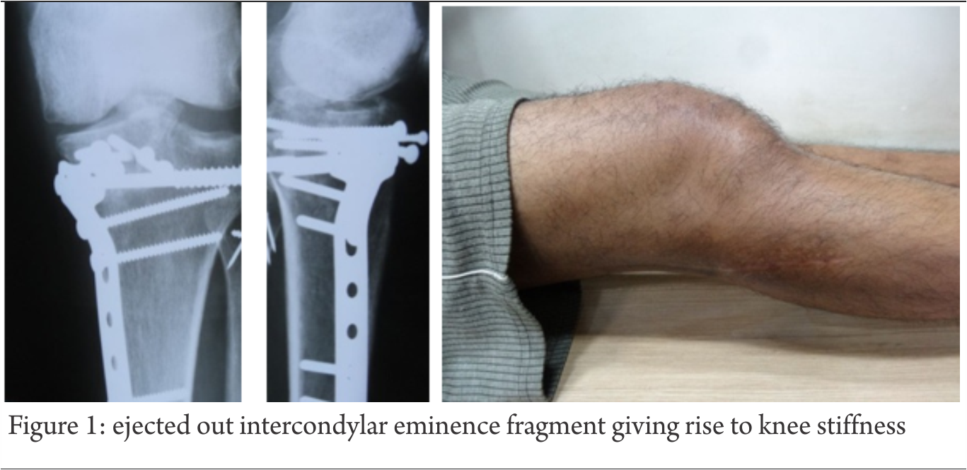
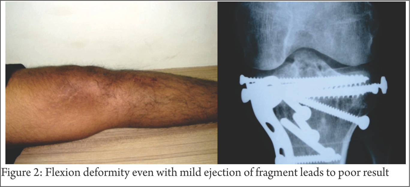
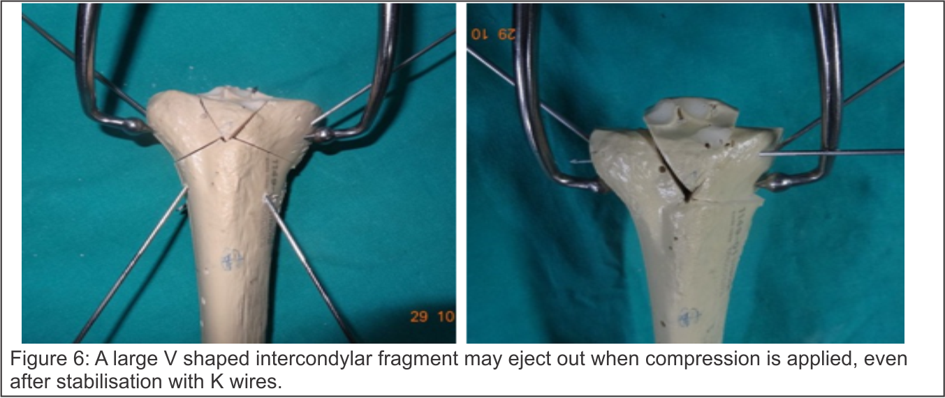
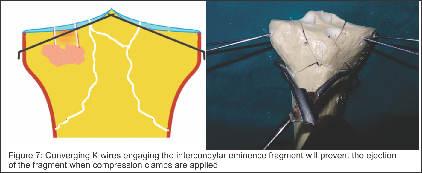
 Volume 2 | Issue 1 | Jan-Apr 2016 | Page 12-16|Majed Al-Mourgi1


Author: Majed Al-Mourgi[1]

Address of Correspondence


Abstract

Purpose: The purpose of this study is to assess the effectiveness and impact of trauma care system, according to the survival in the case of trauma. The main focus of this study is to evaluate the patient survival rates caused by trauma and checking either it is increasing or decreasing due to trauma system. Methods: In this research study, the quantitative data analysis approach is used to examine by the use of different statistical tools. Multiple linear regressions used to find the effect of independent variable on the dependent variable. Results: The results that obtained from the multiple statistical and different important tools must predominantly recognize the effectiveness of trauma system implementation and its impact on patient survival rates. The p-value is found to be significant as it is less than the predefined level of significance. Conclusions: The results defined the effects of trauma care system is used to increase the survival rates. With the development of trauma care center the survival rates is significantly increase and the effectiveness of the trauma care system is depending on the response time taken for trauma injury
Keywords: Patient, Quantitative Research, Survival Rates, Trauma Care System.


Introduction

Trauma is found to be the significant cause of permanent disability and death throughout the world. In USA, a wide study was conducted in order to assess the effectiveness of trauma care system in suburban and urban areas. In the study, patients were originally identified from database of discharge on basis of codes and also stratified according to the severity and type of injury, age and gender [1]. The study provides the detailed results on significant covariates known to cause the risk of death. From 5191enrolled patients were selected in the study from the total of 18,198 trauma patients who are able to meet the inclusion criteria [2]. It is found that the survival rates were significantly increased in patients by getting treatment from trauma care center than in patients getting treatment at a non- trauma care center [3].
According to the nature of trauma, the outcomes of trauma can be improved by developing certain trauma systems which is internationally known as “Trauma Care System” [4]. There are 5000- 6000 trauma patient die according to the report of RTA (Recorded Trauma Accident) [5]. For each person who dies, there are numerous thousand more persons are injured and most of them with permanent sequelae. It is also realized that 48% of the total mortality rate are caused by car accidents. These are the significant increment found in health related problems due to injury throughout the world. From the past history of trauma centre it is observed that every day death of approximately 16,000 people is realized caused by injuries. Approximately 16% of global burden of diseases is caused by injuries [5]. From which less than 90% of the total injuries burden mostly occurs within middle income and low income countries. Another significant cause of mortality in the western countries is patient’s ages from 1 to 44; however trauma strikes at all ages.
The registries of trauma play a significant role in the trauma care system, although the success of such system is hard to compute by means of related official and publications use for quality control. From the report of King Abdullah International Medical Research Center in Saudi Arabia, Trauma is the major sources of suffering, not only cause the patients leading to loss of their life but also cause temporarily or permanently disability in the patients. It is also cause to major economic loss. There is high probability for Trauma found in the young and working age adults, but it is also realized that if injury strikes at geriatric age the mortality ratio is much higher [6].

Background
Inclusive trauma system is found to be the most severe cases which are transferred to a trauma center [6]. In 2006, Utter et al. conducted a study for 2001 severe trauma patients which used inpatient databases in order to identify all severe trauma patients from 24 US states. From these 24 US states, 8 states where classified as ‘exclusive’, also another 8 states were classified as ‘more inclusive’ and the remaining 8 states were classified as ‘most inclusive’. In this study, from the total 61,496 patients with an ISS of 16 or higher were enrolled [13].
After the London bombings, analysis of resource and response studied by Aylwin et al. in 2005. This study showed a test for the trauma care system from the large resource of consumption for a very short period. In this study, researcher realized approximately 775 casualties and total 56 deaths from which approximately 53 were death at the scene. 55 patients were triaged as priority 1 and 2 severely injured. From these 55priority 1 and 2, twenty were critically injured. The realized over- triage rate was 64% and it is also observed that 3 patients died in hospital [14].
In Saudi Arabia, trauma is considered as a major problem related to public health which increases the rates of mortality and morbidity. For the socioeconomic burden, trauma causes the emotional and psychological stress on families, depletion of human resources and the healthcare facilities. In order to minimize these impacts of trauma, a national trauma care system has to be developed and implemented before it is too late to manage the further complexities of trauma in the future.

Research Design
For this study, the research design is used to define the objective of the research study. It is also defined as an effective approach that used to define the nature of research study. Past studies are significantly used to assemble the data in order to identify the nature of research study. The major aim of this research study is to assess the impact of trauma care system and its effectiveness on survival rates. Accidents are the major source of injuries or trauma. Therefore, different descriptive analysis tools are used to evaluate the occurrence of research in order to recognizing and describing the need of competitor analysis [3].

Research Questions
For this study, the research questions are used to explain the problems cause by trauma and implication of trauma care system. Inferential statistical tools are significantly used to solve these problems by collecting and analyzing the secondary data [1].
The primary objective of this population based research study is to evaluate the implication of the trauma system and its impact. For this study, the underlying research problem is to evaluate the effectiveness of the trauma care center with respect to the survival rates. The impact of trauma system for injured patients is found to be a positive sign with respect to previous researches. The major aim of this study is to analyze the impact and effectiveness of trauma care center with respect to the survival rates. For that purpose various descriptive research approaches were used to analyze the occurrence of injuries [11].

· Question 1: Does general team response in the trauma care system play an important role?
· Question 2: Are survival rates significantly increasing by the development of trauma care centers?

Research Hypothesis
In this research study, research hypothesis is used to analyze the stated problem and research design by the uses of the research hypothesis for implication of trauma management. For speculation about the research and experiment, therefore the hypothesis are stated for trauma in order to check the hypothesize research and experiment results [12]. In this study, a systematic approach is developed in a systematic way in which data is collected for the study to address the research question.
For this research study, different statistical approaches are used in order to check the research hypothesis by means of specification, prediction and testable data. The purpose of the state hypothesis is to develop a summary and also to develop the need in operational term and frame work of research [10]. For the study, the hypotheses are stated in order to check the speculation is either confirmed or rejected with the help of the statistical model. The study based on the following hypotheses:

· H1: The general team response does not play an important role of trauma care system.
· H1A: The general team response plays an important role of trauma care system.
· H2: Survival rates are not significantly increasing by the development of trauma care centers.
· H2A: Survival rates are significantly increasing by the development of trauma care centers.

Materials and Methods
Methodology is one of the most significant elements for this research study because author wants to analyze the effect of trauma care system with the help of systematic approach of research. The methodology of a research study is used to draw a design by which the study received help in executing the research. It also provides helped to complete information in an organized manner and realized proper flow to gather all the necessary information which can help to conclude the research hypotheses. Conversely, the research methodology for a specific research study provides a systematic technique in which data is collected and on the basis of collected data research questions can be evaluated or the research hypothesis can be assessed.
In this study, quantitative research methodology is used with an aim to get appropriate outcomes. The research methodology for this study refers to draw the outline in a systematic way in which the data is collected for the study with an aim to provide the valid conclusion about the research questions [6]. It can be more simply defined as the research methodology is a comprehensive plan that incorporates such procedures in order to formulate and state the research question to check the hypothesis [1].
This study is conducted with an aim to get positive increment in the survival rate after implementing trauma care system. Its effectiveness evaluated from the population based study. This study used trauma care center data which is collected throughout the year on monthly bases. The responses provided by the patients are on their traumatic experiences are collected as the quantitative data. The collected data is used to investigate by using multiple statistical techniques that provides appropriate results [12].

Data Collection
In this research study, the secondary quantitative data is used to analyze the research problem. The quantitative data analysis used to examine the effectiveness of trauma system implementation and its impact on patient survival rates. For that purpose the selected data is explained on monthly bases.
The quantitative approach based research study gives serious solutions that are important to identify the main problems of research [9]. In order to estimate appropriate results, the quantitative research is used to identify the importance of statistical significant and contract with numerical data. It totally informs the practical values of various theories that define numerous structures.
Results
Correlation
In statistics, correlation is a technique that is used to show whether and how strongly set of variables are associated with each others. Conversely, it is used to check the effect of one variable on the other variable as increase in one unit how it is affected to other variable. Correlation is defined as the degree by which two variables for the same group of elements explains a tendency to differ to each other [5].
From the outcome of table 1, the strong and positive association found between the team general response and other variables. However, there is a moderate association between the team general response and gastric tube but it is also found that the result is insignificant because the p value is greater than the predefined level of significance i.e., 0. 05.

Regression
In this study, the regression analysis tool is used in order to estimate the relationship between the variables. It is also used to analyze and forecast the post value of general team response in the trauma system on the base of past values [7].
From table 2, the model summary explains that the value of R square is near to 1 which tells us that the fitted linear regression model is accurately stated.
From table 3, the regression coefficient explains the linear relationship between the dependent and independent variable. From the table, there is a negative association found between the dependent variable and independent variable. All estimated results are significant on the bases of p value.

ANOVA
ANOVA (analysis of variance) is defined as the collection of statistical models in order to analyze the association procedure and difference between the groups means [4].
From table 4, the F stat is 779. 325 and the p value is less than the predefined level of significance therefore, the null hypotheses are rejected and conclude that the general team response for the trauma in the trauma care system play an important role and survival rates are significantly increasing by the development of trauma care centers.

Discussion
Trauma has been considered as the most leading cause of injuries and mortality among individuals particularly to those with the age below 45 years. According to this research study, the effectiveness of the trauma care system in hospitals is highly depending on the response of team. It has been observed from the above study that the excessive variables are highly effectual in defining the response time to operate trauma in the trauma care system and its impact for increasing survival rates. The correlation table portrays the positive impact of the response time because of the strong positive strong correlation realized among the selected variables. The results showed that all of these variables were positively associated with the effective team management. Also, the P value is less than the predefined level of significance i.e., 0. 05, therefore the results are significant [8]. The obtained results and statistical outcomes defined on the bases of A&E other roles and functions, Gastric Tube, Hypo/Hyper Thermia machine, Diagnostics -Radiology, Initial Vital Signs, Diagnostics -Lab, Diagnostics Fast, Diagnostics CT Scan.

Conclusion
From the above research results, according to the obtained results the increment of survival rate trauma care system plays a significant role. The study found that for severe injury case there is a very small probability of survival. The goals developed by the mutual and developed from research methodology, there are significant relationship found between the variables. In this study, the effectiveness after implementing trauma care system is realized and also its impact on patient survival rates are also found significant. From the research results there is a significance found in the effectiveness and impact of trauma system in increasing the survival rates. The different variables were analyzed to conclude the hypothesis and study provide the enough evidence to conclude that trauma care centers is significantly increasing the survival rates. The study also concludes that the early response from the team is highly effective for the trauma care system.


References

1. Barisa, M. T. , Dahdah, M. N. , Schmidt, K. , Barnes, S. A. , Dubiel, R. , Dunklin, C. , . . . & Shafi, S. . Comparative effectiveness of traumatic brain injury rehabilitation: differential outcomes across TBI model systems centers. The Journal of head trauma rehabilitation, 29(5), (2014), 451- 459.
2. Lu, M. , Althausen, P. L. , Thomas, K. C. , Shannon, S. F. , Biagi, B. N. , & Boyden, E. M. . Implant standardization for hemiarthroplasty: implementation of a pricing matrix system at a level II community based trauma system. The Journal of arthroplasty, 29(4), (2014), 781- 785.
3. Unsworth, A. , Curtis, K. , & Asha, S. E. . Treatments for blunt chest trauma and their impact on patient outcomes and health service delivery. Scandinavian Journal of Trauma, Resuscitation and Emergency Medicine,23(1), (2015), 17.
4. Porter, A. , Wyrick, D. , Bowman, S. M. , Recicar, J. , & Maxson, R. T. . The effectiveness of a statewide trauma call center in reducing time to definitive care for severely injured patients. Journal of trauma and acute care surgery, 76(4), (2014), 907- 912.
5. Cole, E. , Davenport, R. , Willett, K. , & Brohi, K. . Tranexamic Acid Use in Severely Injured Civilian Patients and the Effects on Outcomes: A Prospective Cohort Study. Annals of surgery, 261(2), (2015), 390- 394.
6. Bodnar, D. , Rashford, S. , Hurn, C. , Quinn, J. , Parker, L. , Isoardi, K. , . . . & Clarke, B. . Characteristics and outcomes of patients administered blood in the prehospital environment by a road based trauma response team. Emergency Medicine Journal, 31(7), (2014), 583- 588.
7. Church, E. C. , Selassie, A. W. , Cao, Y. , Saunders, L. L. , & Krause, J. . Accelerated death rate in population- based cohort of persons with traumatic brain injury. The Journal of head trauma rehabilitation, 29(3), (2014), E8- E19.
8. Bulger, E. M. , Fox, E. E. , del Junco, D. J. , Holcomb, J. B. , Brasel, K. J. , Hoyt, D. B. , . . . & ROC Investigators. (2015). Collider bias in trauma comparative effectiveness research: The stratification blues for systematic reviews. Injury. (2015).
9. Lansink, K. W. , & Leenen, L. P. . Do designated trauma systems improve outcome?. Current opinion in critical care, 13(6), (2007), 686- 690.
10. Yeung, H. H. , Rainer, T. H. , Gabbe, B. J. , Yuen, K. Y. , Ho, H. F. , Kam, C. W. , . . . & Graham, C. A. . A Comparison of Functional Outcome in Patients Sustaining Major Trauma: A Multicentre, Prospective, International Study. PloS one, 9(8), (2014), e103396.
11. Chiu, Y. L. , Allen, B. B. , Gerber, L. M. , Ghajar, J. , & Greenfield, J. P. . Age- specific cerebral perfusion pressure thresholds and survival in children and adolescents with severe traumatic brain injury. Pediatric critical care medicine: a journal of the Society of Critical Care Medicine and the World Federation of Pediatric Intensive and Critical Care Societies, 15(1), (2014), 62.
12. Jurkovich, G. J. . Focusing on the New Reality in Trauma Care. Annals of surgery, 260(1), (2014), 22.
13. Utter, Garth H. , Ronald V. Maier, Frederick P. Rivara, Charles N. Mock, Gregory J. Jurkovich, and Avery B. Nathens. Inclusive trauma systems: do they improve triage or outcomes of the severely injured?. Journal of Trauma and Acute Care Surgery 60, no. 3 (2006): 529-537.
14. Aylwin, Christopher J. , Thomas C. Konig, Nora W. Brennan, Peter J. Shirley, Gareth Davies, Michael S. Walsh, and Karim Brohi. Reduction in critical mortality in urban mass casualty incidents: analysis of triage, surge, and resource use after the London bombings on July 7, 2005. The Lancet 368, no. 9554 (2007): 2219-2225


How to Cite this article:.Al-Mourgi M. Effectiveness of Trauma System implementation and its impact on Patient Survival Rates. Trauma International Oct-Dec 2015;1(2):12-16.

Authors


(Abstract)      (Full Text HTML)      (Download PDF)


Intramedullary Fibula with Rigid Osteosynthesis in Revision of Neglected & multiple times Operated Non Union of Long Bones

 Volume 2 | Issue 1 | Jan-Apr 2016 | Page 12-16|


Author: 

Address of Correspondence


Abstract

Background: Plates with Intramedullary Fibula as Strut graft(IFSG) in Non- union of long bone fractures provide most stable construct overall as fibula acts as second implant, screws have better anchorage and Very High Pull Out strength due to penetration of four cortices and Osteogenic property of the fibula is most helpful.
Aims: re-focus the importance of Non-vascularised intramedullary fibula as a second internal biologic stable splint along with rigid osteosynthesis for the treatment of difficult & neglected non -union of long bones.
Methods and Material:
15 cases (13 :2,M f) of revision surgery for the neglected & multiply operated non-union of the long bones were treated with the debridement, decortication & shingling of the fracture site with addition of the trimmed fibula (as strut) and addition of profuse bone grafting sub periosteally followed by Stable & rigid Internal Osteosynthesis by using LCP. DASH Score was used for upper limb & VAS for lower limb for assessment.
Results: Union was achieved in all patients in 12 months (Av.9-12 Months). Preoperative DASH score averaged 40.86(range 42.6-52.6). DASH score at the last follow-up averaged 20.38 points (range 16.4-24.2).difference was significant. (p=0.0001)
Conclusions: Intra medullary fibula almost works mimicking double plate & adds in the stability as it works as second plate & osteogenic property helps in the healing, not only at the fracture site but proximal & distal to it also thus avoiding SLOW-UNION at the ends of bone which are usually avascular because of the lysis. Screws have a better hold around osteoporotic bones due to four cortices hold in presence of fibula. Strong bony union can be successfully achieved in almost all cases with rigid compression at the fracture site & additional extensive cancelleous bone grafting.
Key-words: Intra- medullary Fibula ,revision Non Union,rigid Osteosynthesis


Introduction

Non -union, of Multiple times operated Long Bone fractures, poses the extreme challenge to the orthopaedic surgeons
Surgical treatment of proxiamal humeral non unions and malunions are technically challenging. Osteosynthesis with bone grafting for the treatment of nonunions is indicated in young, active patients with adequate bone stock in the proximal fragment and preservation of the glenohumeral articular surfaces[2]
Vascularised bone grafting requires surgical experience and equipment not readily available in every hospital. The technique is demanding of time and resources, and vascular thrombosis may compromise the result [5]

Iliac crest cancellous bone graft has no mechanical strength to withstand stresses prior to solid union of the fracture site and also is associated with quite significant graft harvest site morbidity. The fibula transmits one eighth of body weight and can be as useful as vascularised or non-vascularised graft in reconstruction of bony defects.

Compression plating with autogenous grafting is accepted as the gold standard method so has yielded satisfac¬tory results, with 92 to 100% healing rates
Operative treatment can be very successful when the techniques of plate-and-screw fixation are modified to address osteopenia and relative or absolute loss of bone. Healing of the fracture substantially improves function and the degree of independence[6]

Subjects and Methods: 15 patients of multiply operated non union of long bones (13 humerus, 1 femur and one distal tibia non union) were included in a study. All were treated by open reduction of the fracture, debridement and excision of the fibrosis, SHINGLING of the bone both proximally and distal to fracture site.
& osteosynthesis with strut non vascularised fibular graft and LOCKING COMPRESSION PLATE with few locking screws and rest of all are cortical screws to make it RIGID & STABLE CONSTRUCT with extensive cancelleous bone grafting 3600 surrounding the fracture site.
All patients were assessed at final follow up using D.A.S.H score for upper limb and Visual analogue score for lower limb.
All patients were followed up for 4 to 36 (average mean months is 20 months) except two patient, who are under study with last two-two and half months.
Out of 15 patients,13 were male and 2 were female. Out of 15, 13 patients had humerus non union,two had femoral mid shaft non union and remaining one had lower third distal tibia non union.
Out of the 13 humerus cases 4 had proximal humerus, 7 had mid shaft to lower third humerus(M3-L3RD) non union & two patient has non union of segmental shaft humerus fracture.
All were closed fractures except one open fracture and all had eventual atrophic non-union upon presentation. The patients had mild pain, tenderness and abnormal mobility at the non-union site (except distal tibia and middle femur), and limitation of activities of daily living. All patients had stiff¬ness of shoulder and elbow to varying degrees.
The patients with femoral non unions have constant pain at the fracture site and difficulty while bearing weight.
distal tibia non union patient had not started bearing weight as he was on fixator for three months following the Gun shot injury operated in other country.

Surgical technique:
We discussed, here (figure 1 A,1B) is a case of 52 years old male, who was operated 4 times within last 10 years & presented to us with this latest x rays showing atrophic gap Non union of the Proximal humerus.

Fracture site is exposed using previous scar in all cases as to avoid cosmetic ugly scar. The fibrous non-union and any devitalised bone were thoroughly excised and the medullary canal opened via sharp thinner humerus awl and 3.5-4.5 mm drill bits.
• 3. Preparation of the medullary canal: The humeral medullary canal was prepared to accept the bone graft. Fibrous and pseu¬doarthrosis tissue were removed completely and the medullary cavity reconstituted both proximally and distally by curettage, drill or the use of serial hand reamers (6mm-9mm). Dilate humerus medullary cavity mainly to measure the cavity
• Uniformly expanded medullary cavity by the reamers was prepared to put in the graft.(FIGU 2A,2B)
• 4. preparation of fibular graft: The mid-shaft of the fibula was then harvested under tourniquet control with care taken to identify and protect the superficial peroneal nerve. The fibular shaft of ex¬cess length was harvested so that it could be trimmed as neces¬sary. The fibula graft was trimmed so as to enable it to telescope snugly into the fragments across the fracture site. Size of fibula graft will be one smaller than Last reamer used.
If the thickness did not permit its use, it was conversely bevelled at one of the ends leaving behind proximal wider part in hollow cavity of proximal humerus and remaining part in the distal shaft. Generally in humerus ,it necessary to split fibula in the center, with oscillating saw or reciprocating saw or large bone cutter instruments to reduce the size (FIG 3C & 3D)
5. Insertion of the fibular graft: The fibular autograft was pushed into one of the fracture fragments and the exact length of graft that need¬ed trimming was assessed. Once the final shaping of the graft was done, the fracture was reduced with the intramedullary fibu¬lar graft spanning the fracture site. Confirm it is movable in the humerus medullary cavity on both sides of # easily.
• Plate fixation: Reconfirm graft moving in canal push it up all the way proximally(fig 5 A,B,C& D)
• Reduce # , distract ,hold fibula end and slowly push it distally(fig 4A,4B)
• Compress # maximally. Load cancellous graft after shingling (Fig 4 c & 4 D)

How to achieve compression at the fracture with intramedullary graft:
It is safer to do compression by Muller’s device as it is applied at the end of the bone and maximum compression can be achieved by this device. If one chooses to compress by D C P holes. Fix screw on one side of fracture , and for opposite side D C P hole should be used which is not going thru the fibula , as if fibula is fixed it will not allow compression. So under C arm see the end of fibula and go beyond it to put other D C P screw and compress . Generally one hole compression is not enough in this non union set up, so it will have to be reapeted also on 2nd hole . so it is suggested to use muller’s device or a webers device to do compression of the fracture.

A LCP was used with quadricortical screw fixation till fibula is extending. Each screw hole was drilled and tapped through four cortices, two in the fractured bone and two in the intact fib¬ula and 4. 5 mm cortical screws were inserted. End of the bone screws were from parent bone and few locking screws were used for osteoporosis.

Shoulder and elbow exercises were started a day after the opera¬tion. Lifting of weights using the operated limb was deferred for a period of three to four months or until osteo-integration of the fibular ends or fracture healing. After hospital discharge, patients were observed on a monthly basis until healing of the fracture. All pa¬tients were examined both clinically and radiologically. Fracture union was considered radiographically if callus formation was seen in three of four cortices on AP and lateral views. Clinical union was considered when the fracture site was painless.
Data collected retrospectively included grip strength, range of motion, radiographic parameters, and functional outcomes as measured by the DASH (Disabilities of the Arm, Shoulder and Hand) questionnaire.

Results: Patients were followed-up for an average of 1.5-2 years (range 1-2 years). Union was achieved in all patients in 12 months (range9-12 months ) and patients were very satisfied with the treatment. There were no perioperative complications such as wound infection, radial nerve palsy, hematoma formation. Post op two patient had discomfort over the fibular graft harvest site, and one has ASIS graft site morbidity in the form of pain mainly. One patient has persistent fracture line visible on fresh x ray at the shaft of humerus at one year follow up, but still fracture seems to be “ CLINICALLY UNITED “ as patient has painless arm movement.

Preoperative DASH score averaged 40.86(range 42.6-52.6). DASH score at the last follow-up averaged 20.38 points (range 16.4-24.2).(p=0.0001)[Table 1] V.A.S (visual analogue scale) in one patient was improved from 6-7 to 2 at the last follow up.[table 1] There was an average loss of 10-20˚ abduction and 15-20˚ flexion of the shoulder. . Range of motion of adjacent joint (knee in lower limb and elbow in upper limb) was restricted in all patients because of history of multiple surgeries and immobilization periods, but was within the functional limit in all except two patients. There was no change in shoulder rotations following surgery on the contrary three patient had pain free rotation of shoulder who had atrophic proximal humerus non union. One patient with preoperative fixed flexion deformity of elbow to varying degrees had persistence of a similar deformity at the last follow-up.

Discussion: Non -union of the long bone, after repeated surgery with multiple failed attempts poses challenging reoperation.
Healing is challenge & unpredicted with any kind of surgery and may have sub-optimal result .
We have done 15 cases of previously operated multiple times with failed union
As we have used massive mixed cortico-cancelleous auto graft in almost all cases circumferentially at the non-union site –the reason why it unites in each cases is not definite what worked out of these procedure is not predictable, like shingling & solid compression at the fracture site+cancelleous autograft Vs IMSF autograft.
Before we conclude, that fracture is united, we need evidence of circumferential callus formation , and osteo-integration, which takes long time , to be observed . Though patient is using arm almost normally, probably due to good long bony rigid fixation, and hence clinical signs of union are not convincing. This is observed most often in such multiple times operated cases only after 12 months and so we feel, 12 months minimum time should be considered for union.
The main weakness of our study is
1. Very few number of cases(only 15 cases)-so difficult to judge trend of results
2. What exactly helped union , out of everything we did.
The main strength of our study:
Our procedure can not only salvage the “function less –atrophic non united bone “—-it can even have profound effect on the overall compliance of the patient, reduce repeated morbidity from the lengthy & costly treatment like illizarov, which has frustrated outcome and at the same time gives pain free extremity function up to its fully maximum ability.
We did not seen any complication like post op infection, radial nerve palsy, implant cut through, not even
fracture of the fibula graft in any case. Two patient had fibula graft site pain, which subsided within 6 months and another female patient had ASIS graft site pain mainly which also settled within a year.
The reason for low complications in all cases may be we were lucky .
Osteoporosis, either as a result of disuse or due to
generalised metabolic causes, compounds the choice of surgical treatment in these patients. It significantly reduces the pull out strength of the screws thus increasing the chance of implant failure. Humeral nonunion in osteoporotic bone presents a reconstructive challenge for the treating orthopaedic
surgeon [3,4].
In case of non-union, mechanical stability at the fracture site and biologic re¬vitalization are keys for the management.
A non-union of a diaphyseal fracture of the humerus can present a major functional problem even in the elderly population. Advances in operative exposures combined with newer techniques of achieving stable internal fixation even in the presence of pathologic bone have enabled the surgeon to successfully treat even the most complex non unions [7]
Approximately 10% of all long-bone fractures occur in the humerus. Although primary treatment usually is successful, humeral nonunion can lead to marked morbidity and functional limitation. Complications include joint contractures of the shoulder and elbow, especially with periarticular pseudarthrosis. Marked osteopenia or bone loss, or both, often occur after fracture and after failure to achieve union. Retained implants often break, impeding fixation and requiring removal. Soft-tissue deficits and incisions from the original injury or prior surgeries also may complicate reconstruction, as can intra-articular fractures and associated nerve palsies. Successful surgical management of humeral nonunion requires stable internal fixation that allows early joint motion and uses autogenous bone graft to promote healing [8,9]
DCP with cancellous bone graft is a reliable and an effective treatment for revision of aseptic nonunion of humeral shaft fracture after surgical treatment [10,11]
We have used LCP in all 15 cases.
.There are four main indications reported in the literature for use of LCP in fractures:1)osteoporoticbone,
2) comminution, 3) intra-articular fracture, and4) short segment periarticular fracture [12,13]
Locked plates and conventional plates rely on completely different mechanical principles to provide fracture fixation and in so doing they provide different biological environments for healing. Locked plates may increasingly be indicated for indirect fracture reduction, diaphyseal/metaphyseal fractures in osteoporotic bone, bridging severely comminuted fractures, and the plating of fractures where anatomical constraints prevent plating on the tension side of the bone. Conventional plates may continue to be the fixation method of choice for periarticular fractures which demand perfect anatomical reduction and to certain types of nonunions which require increased stability for union.[14]

Conclusion:
Non-vascularised intramedullary fibula(along with debridement, decortication & shingling ) as a second internal biologic stable splint along with rigid osteosynthesis can achieve excellent Union for treatment of revision of difficult & neglected non -union of long bones.


References

1.Gwathmey FW Jr, Jones-Quaidoo SM, Kahler D, et al. Distal femoral fractures: current concepts. Re- view. J Am Acad Orthop Surg 2010;18(10):597–607
2.Agarwal A. Open reduction and internal fixation of the distal femur. In: Wiesel SW, editor. Operative techniques in orthopaedic surgery, vol. 1. Philadel- phia: Lippincott Williams & Wilkins; 2011. p. 582–4.
3.Haidukewych GJ. Temporary external fixation for the management of complex intra- and periarticular fractures of the lower extremity. J Orthop Trauma 2002;16(9):678–85.
4.SinghAK,RastogiA,SinghV.Biomechanicalcompar- ison of dynamic condylar screw and locking compres- sion plate fixation in unstable distal femoral fractures: an in vitro study. Indian J Orthop 2013;47(6):615–20.
5.Henderson CE, Lujan TJ, Kuhl LL, et al. 2010 mid- America Orthopaedic Association Physician in Training Award: healing complications are common after locked plating for distal femur fractures. Clin Orthop Relat Res 2011;469(6):1757–65.
6.Stoffel K, Dieter U, Stachowiak G, et al. Biomechanical testing of the LCP–how can stability in locked internal fixators be controlled? Injury 2003;34(Suppl 2):B11–9.
7.Prayson MJ, Datta DK, Marshall MP. Mechanical comparison of endosteal substitution and lateral plate fixation in supracondylar fractures of the femur. J Orthop Trauma 2001;15(2):96–100.
8.Beltran MJ, Gary JL, Collinge CA. Management of distal femur fractures with modern plates and nails: state of the art. J Orthop Trauma 2015;29(4):165–72.
9.Basci O, Karakaslı A, Kumtepe E, et al. Combination of anatomical locking plate and retrograde intrame- dullary nail in distal femoral fractures: comparison of mechanical stability. Eklem Hastalik Cerrahisi 2015; 26(1):21–6.
10.Suk M, Desai P. Supracondylar femur fractures. In: Archdeacon MT, editor. Prevention and manage- ment of common fracture complications. Thorofare (NJ): Slack; 2012. p. 236.


How to Cite this article:. Bagaria V, Shah S, Sharma G. Distal Femoral Fractures: Complications and How to Avoid them?. Trauma International Jan-Apr 2016;1(2):12-16.a

Authors


(Abstract)      (Full Text HTML)      (Download PDF)