How to Avoid local Complications during Primary and Revision surgery of Trochanteric fractures: Advices and Tricks for operation tactics and Trouble-shooting

Vol 1 | Issue 1 | July – Sep 2015 | page: 21-24 | Andreas Heinrich Hugo Tiemann[1], Ralf Herbert Gahr[2] .


Author: Andreas Heinrich Hugo Tiemann[1], Ralf Herbert Gahr[2].

[1]SRH Zentralklinikum Suhl, Medical Director,Albert-Schweitzer-Str. 2 ,98527 Suhl.
[2]Klinikum St. Georg Leipzig, Delitzscher Str. 141, 04129 Leipzig.

Address of Correspondence
Dr Ralf Herbert Gahr
Klinikum St. Georg Leipzig
Delitzscher Str. 141, 04129 Leipzig
Email: ralfgahr@sanktgeorg.de


Abstract

Background: Hip fractures belong to the most frequent fractures of elderly people. The treatment follows a specific algorithm that focuses on the medical as well as the social situation of these patients. Despite modern operative techniques and implants complications may occur. They may be distinguished into two groups: local complications and systemic complications
Especially local complications (cut out of the femoral head screw) are the reason for revision surgery. Surgery (“salvage procedures”) may only be performed after critical analysis of the local situation and individual planning of the revision procedure. This article outlines the tactical course of action in these cases.
Keywords: Hip fractures – complication – salvage procedure – therapeutic algorithm.


Introduction

Hip fractures are typical injuries of the elderly [1]. Recent studies prove that the incidence of these entities rises dramatically with increasing age [1]. In 2008 Lohmann et al. showed an incidence of 0.06 percent at an age between 60 and 64 years and an incidence of 1.3 percent at people at their eighties [2]. In other words: More than 90 percent of the patients are in their seventies and in addition more than 70 percent are females [3].
American authors assume, that the number of hip fractures will increase from 250,000 in the year 1990 up to 500,000 in 2040 [4]. In Germany the rate of hip fractures totals up to 90/100.000 inhabitants for all classes of age but is 966/100.000 for septuagenarians [5, 6]. Till 2050 the incidence is assumed to multiply by five [6].
In case of geriatric patients suffering from a hip fracture leads to a significant decrease of life expectancy accompanied by an increased medical risk and a number of social problems (for example accommodation in a retirement home) [7, 8]. Anglo-American analyses indicate an average reduction of life expectancy of 1.8 years or 25 percent of the remaining life span per patient [9]. The treatment of such injuries demands a specific algorithm that takes the local surgical problems and co-morbidities as well as social necessities into consideration. Highest goal of the surgical treatment is the early mobilization, prevention of secondary complications and the return to maximal autonomy. Hip fractures today are regularly treated by operative stabilization [10]. Nevertheless the ideal operative procedure is still discussed in the literature [11].
Even modern operative techniques and implants these procedures show adverse events or complications in 15 to 20 percent [12, 13]. Based on acuteness, topography and implant association they may be distinguished into the following groups [14]:
A. Acuteness
Early complication (within three months after surgery)
tactical surgical failure
technical surgical failure
local complication (like wound healing disturbance or infection)
systemic complications (like deep vein thrombosis)

Late complication (after more than three months postoperatively)
effect of poor bone biology (like delayed union or pseudarthrosis)
contracture

b.Topography
local complication (see above)
systemic complication (see above)

c. Implant association
implant associated complication (like cutout, peri-implant fracture or implant failure); (see below)
non implant associated complication

(Revision) Surgery: Planning and tactics
In reference to the special situation of aged patients one has to pay attention to two central factors no matter if one performs primary or revision surgery:
· short term operation
· full weight bearing possible after surgery
Thus the planning of surgery must be focused on the prevention of tactical and technical failures. Revision surgery necessary because of local complications may be divided into two groups:
· Implant (osteosynthesis) related procedures.
Planned implant replacement (for example progressive coxarthrosis that leads to removal of an intramedullary stabilization system in order to implant a total hip arthoplasty).
Enforced implant replacement based on local complications (salvage procedures)

Not implant related local surgery (like the relief of a local hematoma).
A strict separation between these two types of complications is not always possible in praxi. Based on the above named principles one has to keep in mind the following general standards [15]:
· On the part of the surgical procedure:
o full weight bearing possible after surgery
o stable implant fixation in osteoporotic bone
o preservation of the vascularization of the femoral neck and head
o simple operative technique

· On the part of the surgeon:
o profound knowledge of possible implants and surgical techniques
o mastery of the intramedullary and extramedullary surgical options as well as of arthroplasties
oclear Nevertheless the different studies still show different results indication for a specific implant.

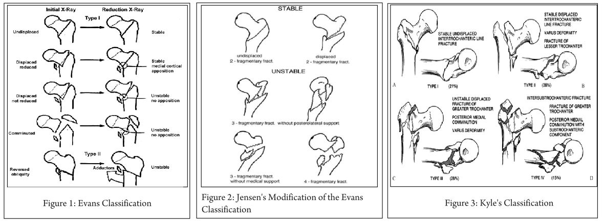
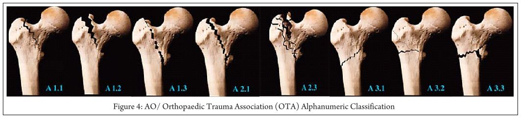
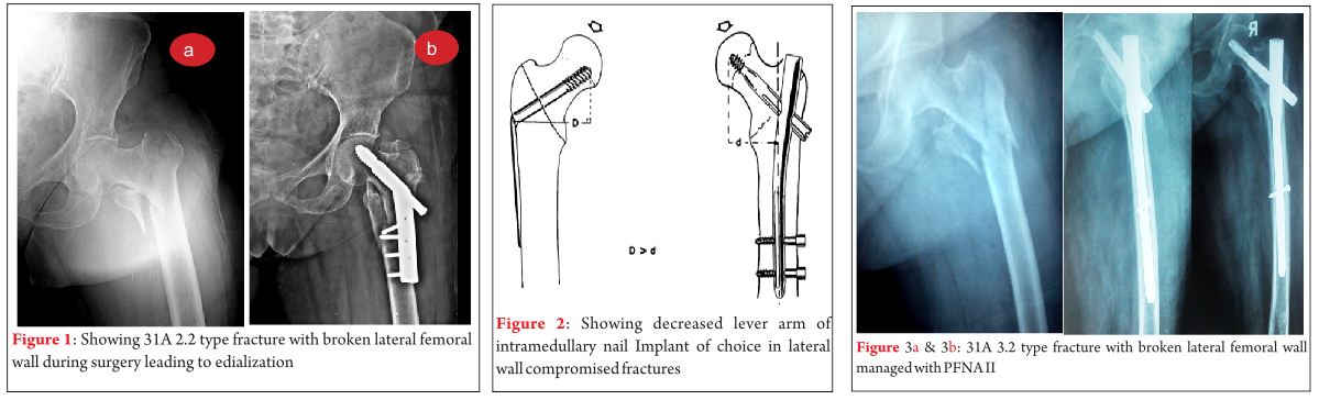
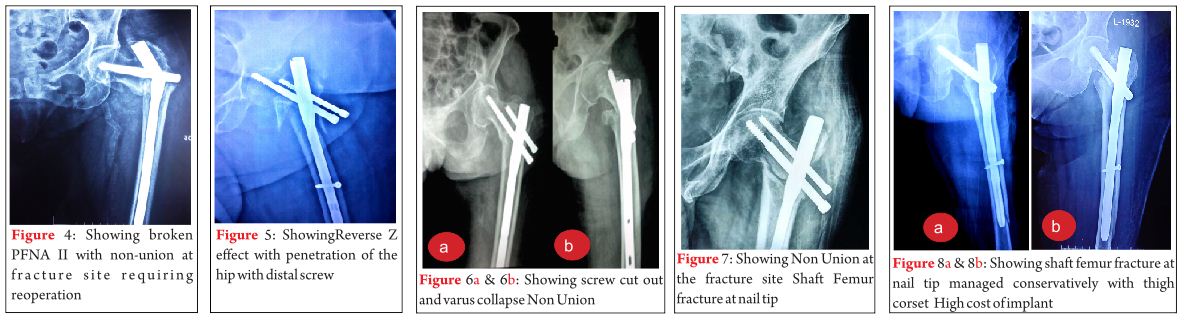
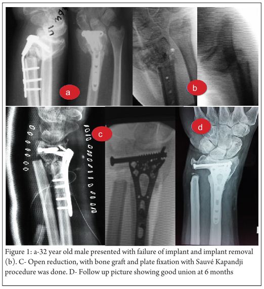
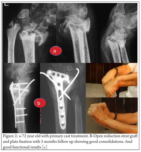
Key-factor I: Preoperative analysis (Fig. 1 and 2)
Primary surgery as well as revision surgery demands a proper analysis of the local situation. The following questions have to be answered preoperatively [15]:
·Where is the problem localized (femoral head and/or femoral neck, trochanteric region, subtrochanteric)?
·Why did the complication occur?
·How may the problem be solved (reosteosynthesis versus (hemi) arthroplasty)?
·What is the correct implant?
·When is the optimal point in time for the operation?

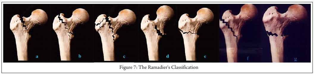
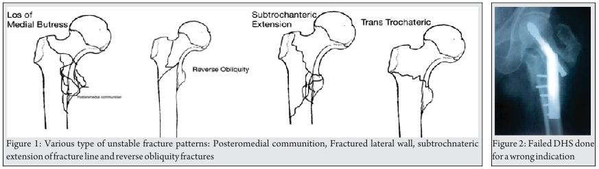
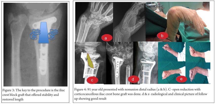
Key-factor II: Osteosynthesis or arthroplasty (Fig. 3)?
In order to answer these questions the following factors are important:
Is the actual situation generally applicable to be treated with an osteosynthesis?
o from the part of the fracture
o from the part of the hip joint (i.e. presence of a significant symptomatic coxathrosis)
If the answer to this question is YES: Which technique will be performed?
o intramedullary stabilization
oextramedullary stabilization
If the answer to this question is NO: Which alternative technique should be performed (hemiarthroplasty, total arthroplasty) [16, 17, 18, 19]?

Stable fractures (AO/OTA 31-A1) present the classic indication for osteosynthesis. The cure rate is close to 100 percent [20]. These fractures mainly are stabilized by the use of a dynamic hip screw (DHS).
AO/OTA A31-A2 and –A3 are unstable fractures. The optimal treatment is still open to debate in the recent literature [16, 17]. Intramedullary stabilization is considered to be better in terms of biomechanical considerations [21]. Nevertheless osteosynthesis failure is estimated to be 56 percent [20].
The question whether to perform reosteosynthesis or switch to hip arthroplasty is elaborated just by few studies. The different studies show different results. Nevertheless Faldini 2002, Sinno 2007 and Giannotti 2013 and 2014 indicate, that elder patients suffering from those unstable pertrochanteric fractures benefit from treatment by cemented hemiarthoplasty. This leads to faster mobilization and lower mortality [16, 17, 18, 19]. According to D`Arrigo et al. the candidates for proximal femur replacement are patients with non-viable proximal femoral segments, destruction of the proximal femoral articular surface, advanced age and the absence of coxarthrosis [22]. Total arthroplasty may be taken into consideration in case of patients with additional severe coxarthrosis.
Switching from internal fixation to arthoplasty offers some special problems [22]:
· failed internal fixation (sometimes accompanied by broken screws) must be removed
· necessity of special instruments in order to remove the failed system
· compromised proximal femur with bone loss distal to the typical neck-resection level for hip arthroplasty

Key-factor III: Open or closed reduction (Table 1)?
The preoperative fracture analysis with a view to it`s reductive capacity based on the radiographic findings (X-ray, CT-scan) is an integral component as well for primary surgery as for revision surgery when reosteosynthesis seams to be possible. Open and closed reduction is the typical approach. Normally AO/OTA 31 fractures will be closed reduced. However between three and 17 percent are estimated to be applicative for closed reduction [23, 24, 25]. In their study Sharma et al. identified 4 general fracture situations, which are not applicative for closed reduction [25]:
· AO/OTA 31-A1 fractures where the proximal fragment is locked underneath the overridden and mediatized shaft fragment
· AO/OTA 31-A1 fractures with bisected lesser trochanter
· AO/OTA 31-A2 fractures with entrapment of the posteromedial fragment at the fracture site
· AO/OTA 31-A2 fractures with an anteriorly displaced proximal fragment and an underlying separate lesser trochanter
The mentioned AO/OTA 31-A1 fractures mainly occurred in younger, the AO/OTA 31-A2 fractures in older patients.

Implant related specification of typical complications of osteosynthesis
In case of planned (re-) osteosynthesis knowledge of the specific osteosynthesis related complications is vital in order to avoid local surgical complications. In general the complication rate is estimated to be three percent [26]. Sathiyakumar et al. distinguished five different treatment variant with significantly different typical implant related complications [9].
The complications mentioned below may occur after intramedullary stabilization as well as extramedullary stabilization of AO/OTA 31 fractures.

Particulars and choice of typical complications of extramedullary systems
The overall complication rate is estimated to be between 6.8 and 16.7 percent [27]. The rate of mechanical complications is estimated to be 2.8 percent [26].
Typical complications
· Cutout. Between three and 16.7 percent for dynamic hip screws [27, 28, 29].
· Z-effect. Mentioned below (intramedullary complications)
· Jamming of the lag screw. According to Simpson this is one of the main reasons for dynamic hip screw failure [30].
· Peri-implant fracture. Parker et al. found it in 0.1 percent in their 2010 study [27].
· Implant overload
· Implant dislocation

Particulars and choice of typical complications of intramedullary systems
The overall complication rate is estimated between 3.6 and 18 percent in the recent literature [26].
Typical complications
·Dislocation of the lesser trochanter
·Varus dislocation of the fracture
·Z-effect-A classical complication of two screw systems (like the proximal femoral nail). The repetitive axial loading of the implant combined with an unstable fracture fixation situation leads to toggling of the nail within the femoral canal [31]. This leads to medial migration of the nail. This mechanism is also described in the 2008 study of Weil et al. [32]. Nevertheless the precise etiology requires further clarification [33].
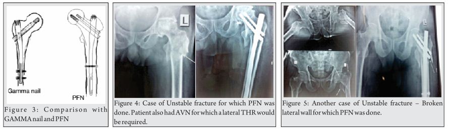
·Cutout. The Cochrane analysis of 2010 describes a rate of 3.4% for intramedullary implants [27]. The contemporary 2014 study of Greorgiannos evidences a rate between two and ten percent [34]. For intramedullary system as well as extramedullary ones the possibility of cutouts depends on the positioning of the lag or sliding screw in the proximal fragment [34]. As early as 1995 Baumgartner et al. described the so-called “tip-apex-distance” (TAD). This nowadays is accepted to be the central principle for the optimal positioning of the lag screw. It is determined intraoperative in the ap. and axial view fluoroscopy [35]. Kuzyk et al. showed in 2012 that the position of the lag screw next to the “Adam´s bow” in the ap. view and in the center of the proximal fragment in the axial view optimizes the biomechanical stability of the osteosynthesis [36]. In addition Nikoloski et al. could prove, that the original TAD rule should be modified depending on the used system, especially when “two screw systems” are used [37]. These authors distinguished two main types of cutout [37]:
o cephaled cutout
o axial cutout
· Knife effect
· Secondary screw dislocation
· Peri-implant fracture. Robinson et al. described an incidence of 18.74 fractures per 1000 person years in case of fractures treated with intramedullary systems [38]. The analysis of Parker et al. in 2010 showed an incidence of 2.6 percent [27]. By developing new designs of the intramedullary devices in the last decades the rate of these complication could be decreased dramatically.
· Implant failure

Discussion

Trochanteric femoral fractures comprise 50 percent of geriatric hip fractures. [39]. Although union rates as high as 100 percent have been reported after primary reduction and internal fixation with stable fractures ideal implants and optimal reduction failure rates over 50 percent are described in case of unstable fractures, suboptimal fracture fixation and poor bone quality [40]. Thus the correct individualized primary stabilization already presents a demanding problem. Appropriate management of failed primary osteosynthesis is even more challenging.
According to the recent literature the choice of treatment depends on the clear identification of the fracture type combined with the specific local situation [39]:
· AO/OTA classification of the fracture.
· radiographic analysis of the local bone structure
o presence or absence of osteoporosis
o presence or absence of extended bone loss
o presence or absence of significant coxarthrosis
When, based on the AO/OTA classification and the above-mentioned facts, the decision for internal (re-) fixation is made optimal reduction of the fracture fragments and positioning of the chosen implant is from central interest [35]. According to the recent literature implant failure generally seems to be the result of poor fracture reduction, mechanical stress, fracture instability or technical error [40] Although the optimal treatment for unstable fracture situations {AO/OTA 31-A2 and A3) are still discussed controversially in the last years a rising number of studies presents good results for arthroplasty as well for primary as for revision surgery [41,42]. Unfortunately there are only few comparative studies and even fewer prospective, random controlled ones which compare prosthetic hip replacement with the standard internal fixation in terms of primary and revision surgery [16]. In their 2014 study these authors come to the conclusion that there are no major differences between the treatment with hemi- or total arthroplasty but especially the elderly patients with severe osteoporosis or comminuted trochanteric fracture take significant profit from the treatment with hip arthroplasty compared to internal fixation [Giannotti]. According to Parker et al. in 2006 there are no significant differences between arthroplasty and internal fixation for mechanical complications, local wound complications, general complications and mortality at one year or long-term function [42].

Conclusion

· proximal hip fracture treatment is no trauma surgery for beginners
· local bone and fracture situation have to be clearly analyzed before taking one or the other surgical option into consideration
· primary stabilization as well as reoperations show specific pitfalls which may affect the outcome of any chosen treatment option significantly
· surgeons have to have profound knowledge about how to identify these pitfalls in order to avoid technical and tactical failure
especially in unstable fracture situations and in case of failed primary internal stabilization arthroplastic treatment should be a serious alternative.


References

1. Prokop A, Chmielnicki M. Pertrochantäre Femurfraktur – eine Anfängeraufgabe? Z Othop Unfall 2014; 152: 584-587
2. Lohmann R, Frerichmann U, Stöckle U et al. Proximal femoral fractures in the elderly. Analysis of data from health insurance providers on more than 23 Million insured persons – Part 1. Unfallchirurg 2007; 110: 603–609
3. Queally JM, Harris E, Handoll HH, Parker MJ. Intramedullary nails for extracapsular hip fractures in adults. Cochrane Database Syst Rev. 2014 Sep12;9:CD004961.
4. Gilbert TB, Hawkes WG, Hebel JR, Hudson JI, Kenzora JE, Zimmerman SI, Felsenthal G, Magaziner J. Spinal anesthesia versus general anesthesia for hip fracture repair: a longitudinal observation of 741 elderly patients during 2-year follow-up. Am J Orthop (Belle Mead NJ). 2000 Jan;29(1):25-35.
5. Bonnaire F et al. Versorgung proximaler Femurfrakturen. Unfallchirurg 2005; 108: 387 – 400
6. Bonnaire F et al. Behandlung von Schenkelhalsfrakturen. Chirurg 2008;: 595 – 611
7. Smektalla R et al. Zur Prognose der Schenkelhalsfraktur. Beurteilung der Mortalität nach Schenkelhalsfraktur durch sektorenübergreifende Datenzusammenführung. Unfallchirurg 2005; 108: 927 – 937
8. Smektalla R et al. Mediale Schenkelhalsfraktur: Einfluss des Versorgungszeitpunktes auf die Ergebnisqualität. Unfallchirurg 2010; 113: 287 – 292
9. Sathiyakumar V, Greenberg SE, Molina CS, Thakore RV, Obremskey WT, Sethi MK. Hip fractures are risky business: an analysis of the NSQIP data. Injury. 2015 Apr;46(4):703-8.
10. Egol KA et al. Perioperative considerations in geriatric patients with hip fracture: What is the evidence? J Orthop Trauma 2009; Vol 23, No 6: 386 – 394
11. Geiger F et al. Trochanteric fractures in the elderly: the influence of primary hip arthroplasty on 1-year mortality. Arch Orthop Trauma Surg 2007; 127: 959 – 966
12. Parker MJ et al. Hemiarthroplasty versus internal fixation for displaced intracapsular hip fractures: a long-term follow-up of a randomized trial. Injury 2010: 41(4): 370 – 373
13. Klinger HM et al. A comparative study of unstable per- and intertrochanteric femoral fractures treated with dynamic hip screw (DHS) and trochanteric bitt press plate vs. proximal femoral nail (PFN). Zentralblatt für Chirurgie 2005; 130(4): 301 -306
14. Huang HK et al. Displaced femoral neck fractures in young adults treated with closed reduction and internal fixation. Orthopedics 2010; 33(12): 873
15. Tiemann AHH, Hofmann GO. Osteosynthesen hüftgelenknaher Frakturen. Trauma und BK 2012;
16. Giannotti S, Bottai V, Dell’Osso G, De Paola G, Bugelli G, Guido G. The hip prosthesis in lateral femur fracture: current concepts and surgical technique. Clin Cases Miner Bone Metab. 2014 Sep;11(3):196-200.
17. Giannotti S, Bottai V, Pini E, Dell’osso G, De Paola G, Guido G. Clinical and surgical approach of severe bone fragility fracture: clinical case of 4 fragility fracture in patient with heavy osteoporosis. Clin Cases Miner Bone Metab. 2013 Jan;10(1):52-5.
18. Sinno K et al. The effectiveness of primary bipolar arthroplasty in treatment of unstable intertrochanteris fractures in elderly patients. N Am J Med Sci 2010; Dec; 2(12): 561 – 568
19. Faldini A. et al. Surgical treatment of intertrochanteric fractures by total hip arthroplasty in old osteoporotic patients. Scientific Exhibit At 69th AAOS annual Meeting 2002: 12 – 19
20. Haidukewych GJ, Israel TA, Berry DJ. Reverse obliquity fractures of the intertrochanteric region of the femur. J Bone Joint Surg Am. 2001 May;83-A(5):643-50.
21. Schipper IB et al. Unstable trochanteric femoral fractures: extramedullary or intramedullary fixation? Review of literature. Injury 2004; Feb; 35(2): 142 – 151
22. D’Arrigo C, Perugia D, Carcangiu A, Monaco E, Speranza A, Ferretti A. Hip arthroplasty for failed treatment of proximal femoral fractures. Int Orthop. 2010 Oct;34(7):939-42.
23. Moehring HD, Nowinski GP, Chapman MW, Voigtlander JP. Irreducible intertrochanteric fractures of the femur. Clin Orthop Relat Res. 1997 Jun;(339):197-9.
24. Chun YS et al. Technique and early results of percutaneous reduction of sagittaly unstable intertrochanteric fractures. Clin Orthop Surg 2011; 3: 217 – 224
25. Sharma B et al. Pertrochanteric fractures (AO/OTA 31-A1 and A2) not amenable to closed reduction: Causes of irreducibility. Injury Int J Care Injured 2014; 45: 1950 – 1957
26. Tsang STJ et al. When does hip surgery fail? Injury Int J Care Injured 2014; 45: 1059 – 1065
27. Parker MJ et al. Gamma and other cephalocondylic intramedullary nails versus extramedullary implants for extracapsular hip fractures. Cochran Library Database System Rev 2010; 9: Dc000093.
28. Nordin S et al. Mechanical failure of dynamic hip screw (DHS) fixation in intertrochanteric fracture of the femur. Med J Malaysia 2001;56 (Suppl D): 12 – 17
29. Huseh KK et al. Risk factors in cutout of sliding hip screws in intertrochanteric fractures: an evaluation of 937 patients. Int Orthop 2010; 34(8): 1273 – 1276
30. Simpson F et al. Sliding hip screws: modes of failure. Injury 1989; 20(4): 227 – 231
31. Takasago T et al. Intrapelvic Migration of the lag screw in intramedullary nailing. Case Reports in Orthopedics 2014; Vol 2014, Article ID 519045;
32.Weil YA, Gardner MJ, Mikhail G, Pierson G, Helfet DL, Lorich DG. Medial migration of intramedullary hip fixation devices: a biomechanical analysis. Arch Orthop Trauma Surg. 2008 Feb;128(2):227-34.
33. Strauss EJ et al. The “Z-effect” phenomenon defined: a laboratory study. J Orthop Res. 2007; 25: 1568-1573.
34. Georgiannos D et al. Complications following treatment of trochanteric fractures with Gamma3 nail: Is the latest version of Gamma nail superior to its predecessor? Surgery Research and Practice 2014; Article ID 143598;
35. Baumgaertner MR et al. The value of the tip-apex distance in predicting failure of fixation of peritrochanteric fractures of the hip. JBJS Am 1995; vol 77, no 7: 1058 – 1064
36. Kuzyk PR, Zdero R, Shah S, Olsen M, Waddell JP, Schemitsch EH. Femoral head lag screw position for cephalomedullary nails: a biomechanical analysis. J Orthop Trauma. 2012 Jul;26(7):414-21.
37. Nikoloski AN, Osbrough AL, Yates PJ. Should the tip-apex distance (TAD) rule be modified for the proximal femoral nail antirotation (PFNA)? A retrospective study. J Orthop Surg Res. 2013 Oct 17;8:35.
38. Robinson CM, Adams CI, Craig M, Doward W, Clarke MC, Auld J. Implant-related fractures of the femur following hip fracture surgery. J Bone Joint Surg Am. 2002 Jul;84-A(7):1116-22.
39. Murphy DK, Randell T, Brennan KL, Probe RA, Brennan ML. Treatment and displacement affect the reoperation rate for femoral neck fracture. Clin Orthop Relat Res. 2013 Aug;471(8):2691-702.
40. Carpintero P, Caeiro JR, Carpintero R, Morales A, Silva S, Mesa M. Complications of hip fractures: A review. World J Orthop. 2014 Sep 18;5(4):402-11.
41. Kiran Kumar GN et al. Bipolar Hemiarthroplasty in Unstable Intertrochanteric Fractures in elderly: A Prospective Study. J Clin Diagn Res. 2013 Aug; 7(8): 1669-1671
42. Sancheti Kh, Sancheti P, Shyam A, Patil S, Dhariwal Q, Joshi R. Primary hemiarthroplasty for unstable osteoporotic intertrochanteric fractures in the elderly: A retrospective case series. Indian J Orthop. 2010 Oct;44(4):428-3.
43. Parker MJ, Handoll HH. Replacement arthroplasty versus internal fixation for extracapsular hip fractures in adults. Cochrane Database Syst Rev. 2006 Apr 19;(2):CD000086.


How to Cite this article: Tiemann A H H, Gahr R H. How to Avoid local Complications during primary and revision surgery of Trochanteric fractures: Advices and Tricks for operation tactics and Trouble-shooting. Trauma International July-Sep 2015;1(1):21-24.

 Photo


(Abstract)      (Full Text HTML)      (Download PDF)


Management Options and Treatment Algorithm in Intertrochanteric Fractures

Vol 1 | Issue 1 | July – Sep 2015 | page: 12-16 | Anoop C Dhamangaonkar [1]


Author: Anoop C Dhamangaonkar [1].

[1]Lokmanya Tilak Municipal Medical College and Lokmanya Tilak Municipal General Hospital,
Sion, Mumbai- 400022, India.

Address of Correspondence
Dr. Anoop C Dhamangaonkar
2/28,Madhavi Soc., Mogal Lane, Matunga (W), Mumbai-400016.India.
Email: anoopd_7@yahoo.com


Abstract

Background: Intertrochanteric (IT) fractures are one of the most common lower limb fractures. There are many varied treatment options to treat the same. Also, our basic understanding of IT fractures is improving leading to evolution of newer concepts which in turn is leading to designing of newer implants. In this sea of newer treatment options, are all newer implants merely market driven? The new- age orthopaedic surgeon needs to be made aware of the newer concepts of IT fractures, implant designs and also a comprehensive literature support of the various treatment options available like concept of unstable IT farcture, the dynamic hip screw (DHS) system, Medoff’s plate, DHS with a trochanteric stabilization plate, 95 degree Dynamic Condylar Screw, first to third generations of gamma nails, proximal femoral nails (PFN), expandable PFN, InterTAN with integrated interlocking screws, proximal femoral locking plates and use of bipolar hemiarthroplasty. This article also aims to present a practical treatment algorithm for the treatment of IT fractures, but this may not be generalizable to all orthopaedic surgeons.
Keywords: Intertrochanteric; unstable; DHS; PFN; Gamma; hemiarthroplasty.


Introduction

Intertrochanteric (IT) fractures are disabling injuries that most commonly affect the elderly population and also in young. The incidence has increased significantly during recent years due to the advancing age of the world’s population. These fractures were managed by conservative methods before 1930s when the age of fixation began. Through this article we wish to present the gradual yet sudden transition in various treatment modalities.
Till the third decade of the twentieth century trochanteric fractures were treated conservatively. Conservative treatment regimes included, simple support with pillows or splinting to the opposite limb, Buck’s (skin) traction, Well-leg traction, plaster spica immobilization, Russell’s balanced traction and skeletal traction through the lower femur or upper tibia. Nonsurgical treatment of intertrochanteric hip fractures is not preferred but is usually reserved for patients with comorbidities who are not fit for anesthesia. Mortality after non-operative treatment is commonly due to cardiopulmonary complications, thromboembolism, and sepsis [1]. In 1989, Hornby et al compared the nonsurgical treatment with a dynamic hip screw (DHS) in 106 patients with intertrochanteric hip fracture [2]. There was no significant difference in complications, 6-month mortality, pain, leg swelling, or pressure sores. SHS group gave better anatomic reduction and a shorter hospital stay. Patients treated conservatively had greater loss of independence at 6-month follow-up. The authors recommended surgical treatment for medically stable patients. A 1981 prospective trial of 150 patients compared nonsurgical treatment (ie, skeletal traction with a tibial pin) with surgical treatment [3]. The authors concluded that excellent results with traction alone could be obtained provided a good nursing care was maintained with careful attention to bedside physical therapy, respiratory care, deep vein thrombosis prophylaxis, and prevention of ulcers. A 2003 retrospective study reviewed a population database to compare mortality rates in patients with severe comorbidities who were treated either nonsurgically or surgically for intertrochanteric hip fracture [4]. It concluded that the mortality rate was low in the conservatively treated patients provided the patients were mobilized early out of bed to chair. The evidence-based literature supports surgical fixation while also providing valuable information in regard to medically unstable patients who must be treated nonsurgically [2-4].

Operative treatment options
Era of Dynamic Hip Screw (DHS)
During the early 1950s when the use of Smith Peterson nail and Jewett nail-plate was very common, W. Schumpelick et al. revolutionized treatment of IT fractures by presenting the results of the sliding neck screw of the DHS in 1955 [5]. Here any force acting on the femoral head in the direction of the body axis will be diverted by the sliding neck screw into the direction of the axis of the screw, so that it strikes the fracture line approximately at a right angle and acts not as a shearing but as a compressing force stimulating callus formation. Here too varus collapse, shortening, superior cut-out occurred. But in non-union was rare.

What is an unstable intertrochanteric fracture?
Stable intertrochanteric fracture implies that the reduction is stable with a lesser tendency to collapse further post-operatively. Whereas the unstable IT fractures have a tendency to collapse much more post-operatively, especially in varus and retroversion. Fractures with a large posteromedial void, reverse oblique fracture configuration, IT fractures with subtrochanteric extension and IT fractures with a lateral wall fracture [6-10].
Modifications of DHS:
Two major modifications to the DHS are the Medoff’s plate and the DHS with a trochanteric stabilisation plate.
In 1991, Medoff et al used a new axial compression screw plate device for unstable intertrochanteric or proximal subtrochanteric fractures of the hip with no technical failures [11]. The device has an axial compression screw to allow compression along an axis parallel to the femoral shaft. As the fracture settles postoperatively, dynamic axial compression continues. In 1993, Babst et al. stated that the lateralisation of the greater trochanter could be prevented in all cases with a trochanteric buttress plate being added to the sliding screw-plate [12]. This also leads to a limitation of telescoping, with less shortening even with immediate full weight bearing. Leung F et al concluded that the DHS blade plate was an effective fixation option in elderly osteoporotic patients with intertrochanteric fracture femur where the conventional lag screw was replaced with a helical blade [13]. Here, the helical blade leads to better blade fixation in osteorotic bone by impaction of the cancellous bone while insertion rather than destroying bone while reaming for the DHS lag screw.

Other conventional options:
95 degree DCS or angle blade plate is often used to treat the reverse oblique intertrochanteric fractures as they biomechanically mimic the subtrochanteric fractures. Rosso R et al recommend that the DCS may be used to treat subtrochanteric fractures rather than intertrochanteric fractures [14].

Era of Intramedullary nails:
Gamma Nail:
This was one of the earliest intra-medullary (IM) imlants to be designed in 1988. The premise on which the IM implants were designed were:
1. To reduce the varus strain on the implant while weight bearing by reducing the force arm.
2. To provide a lateral buttress with an IM device to prevent the lateralization of the proximal fragment and control the collapse rather than relying on the integrity of the lateral wall.
Bridle et al in 1991, prospectively compared the dynamic hip screw and the gamma nail in100 intertrochanteric fractures in elderly patients [15]. They found no difference in the operating time, blood loss, wound complications, and stay in hospital or the patient’s mobility. But fractures of the femur in 4 cases were seen in the gamma nail group which required further revision surgery.
Leung KS et al in 1992conducted a randomized study in elderly patients comparing gamma nails and dynamic hip screws and concluded similar final outcome with both, but achieved less surgical trauma, less screening time, less blood loss and earlier rehabilitation with gamma nails [16].
Radford PJ et al prospectively compared the Dynamic hip screw and the Gamma nail for fixing 200 peritrochanteric femoral fractures in elderly patients [17]. There was less intraoperative blood loss and a lower rate of wound complications in the patients treated by the Gamma nail. They however had a high incidence of femoral shaft fractures which were related to the implant design. They did not recommend the use of the Gamma nail for these fractures. Curtis MJ in 1994 assessed the rigidity and strength of fixation provided by intramedullary and extramedullar devices for proximal femoral fractures [18]. Stable and unstable intertrochanteric fractures were studied after fixation with Gamma nail and DHS implants. There was no significant difference in the strength of fixation of stable and unstable intertrochanteric fractures between the Gamma nail and the hip screw, although the Gamma nail provided more rigid fixation. In 2001, Adams CI et al prospectively compared the IM nailing with a DHS and side plate in 400 patients [19]. Revision rates, femoral shaft fractures, and lag screw cutout were slightly higher in the IM nailing group but not statistically significant from the DHS cohort. There was no difference in the early or 1 –year functional outcomes. Ahrengart L et al randomized 426 intertrochanteric fractures to be fixed with either the Gamma nail or a compression hip screw [20]. The surgical time was not significantly different between the two groups. In the Gamma nail group, difficulty was encountered with the distal locking technique. The incidence of cephalic position of the compression screw within the femoral head, screw cutout, and intraoperative fracture were higher in the Gamma nail group. Walking ability was the same in both groups. The authors recommended compression hip screws for less comminuted fractures, reserving Gamma nails for comminuted patterns.
Utrilla AL et al compared the Gamma nail with a compression hip screw in 210 stable and unstable fractures and found no difference in total surgical time [21]. However, the Gamma nail group had a significantly lower postoperative transfusion requirement. Mortality, fracture healing, and intra- and postoperative complication rates were not significantly different between the two groups. In patients with unstable fracture patterns, postoperative ambulation was significantly improved in the Gamma nail group.

Newer Gamma nails:
The Preference to the use of Gamma nail for unstable IT fractures after early 2000 was primarily due to the introduction of second generation Gamma nail called the Trochanteric Gamma Nail (TGN) and the third generation Gamma nail called the Gamma3 Nail (G3). Better implant design led to decreased fracture shaft femur, which was the complication seen in early generation Gamma Nail [22].
The G3 proximal diameter is 15.5 mm, 1.5 mm smaller than TGN (17 mm). G3 design also allows distal dynamic locking screw. But the migration resistance in G3 is lesser than TGN. The main reason for this is the smaller diameter in the G3 lag screw measuring 10.5 mm rather than 12 mm in the TGN [22].
The latest edition to the G3 design is the Gamma3Rotational Control, with the addition of a spreading U-Clip over the cephalic screw which increases the surface area and also the resistance to failure [22].
Clinical studies report better results with G3. Varela-Egocheaga JR et al reported 2 cases of cutout and 1 screw protrusion in 40 patients (7.5%) in a series comparing G3 with an extramedullary device [23]. Andruszkow H et al reported that a TAD should not exceed 25mm and a valgus reduction reduces the chances of screw cut-out [24]. de Grave PW et al [25] reported 2 cases of mechanical failure in 61 patients treated with a Gamma3 nail, and Westacott D and Bould M [26] reported no case of mechanical failure in 36 unstable proximal femoral fractures treated with a long Gamma3 nail. Mingo-Robinet J et al stated that there was no relationship between cutout rate and TAD in TGN and G3 [22]. Though they concluded that Gamma3 Nail has higher cutout rates than TGN in unstable fractures.

Proximal Femoral Nail
In 1996, AO/ASIF developed the proximal femoral nail (PFN) as an intramedullary device for the treatment of unstable per, inter and subtrochanteric femoral fractures.
Pajarinen J et al compared the DHS with a proximal femoral nail (PFN) in 108 patients and the main outcome measure was recovery of ambulation [27]. The patients treated with IM devices had a significantly faster return to preoperative ambulation levels. Nuber S et al evaluated 129 patients with unstable intertrochanteric fractures treated with either a DHS or a PFN [28]. Revision rates were similar between the two groups. However, there was a significantly shorter surgical time, shorter hospital stay, earlier full weight bearing and lesser pain intensity at 6-months post-op in the PFN cohort. Zhang K et al stated in a meta-analysis of six studies including 669 intertrochanteric fractures that the PFN group had significantly less operative time, intraoperative blood loss and length of incision than the DHS group [29]. There was no significant difference in the postoperative infection rate, lag screw cut-out rate, or reoperation rate between the PFN and DHS groups. They concluded stating that PFN is a better implant than DHS.

Newer PFN designs
PFNA-II is the modified PFN design meant to match the Asian proximal femoral morphometry. Expandable PFN was designed to retain the mechanical characteristics of a large-diameter nail, to provide the good torsional stability between the femoral neck and shaft obtained by an expendable peg inserted in the femoral head (especially in cases of poor bone quality), and to avoid the need for interlocking screws. However its long term utility needs further literature support. Elis J et al concluded that the expandable PFN was at least as good as the 95 degree DCS after comparing the EPFN and the 95 degree DCS [30]. Jin YM et al concluded that expandable PFN are better implants than DHS and anatomic plates in treating intertrochanteric fractures [31].

InterTAN
This is a newer intra-medullary fixation device which uses proximal intergrated interlocking screws in a figure of eight position to attain compression of the intertrochanteric fracture site.
Wang Q et al compared the InterTAN and the DHS and concluded that InterTAN significantly reduces the operative time, intra-operative radiation exposure, intra-operative blood loss and postoperative complications [32]. The InterTAN can be used in elderly osteoporotic patients and in and unstable intertrochanteric fractures.
Jiang Y et al stated that a proper pre-operative imaging, reaming is essential before inserting the InterTAN [33]. But an open reduction and internal fixation with DHS be preferred in cases where the reduction is difficult.
Nüchtern JV et al concluded that the InterTAN with two integrated screws was able to withstand higher loads than the Gamma 3 nail [34].

Newer Proximal femoral Locking plates:
Many new designs of proximal femoral locking plates have been used to treat intertrochanteric fractures. But there is not enough literature evidence of they being better than IM implants. Haq RU et al concluded that PFN is a better implant than reverse-distal femoral locking plate for intertrochanteric fractures with a deficient lateral wall [35]. Zhong B et al stated that the proximal femoral locking plates were better for treating subtrochanteric fractures rather than intertrochanteric fractures [36].
Azboy I et al concluded that both proximal femoral locking plate and 95 degree angle blade plate gave equally good results in reverse oblique intertrochanteric fractures. However, angle blade plates were preferred as they were an economical option [37].
Dhamangaonkar AC et al compared the Proximal femoral locking plate with the DHS. They concluded that proximal femoral locking plates decreased the chances of limb shortening and medialisation of the shaft [38].
A recent study by Shen J et al concluded that these plates are an effective and safe method in the treatment of all types of intertrochanteric femoral fractures, but good fracture reduction and ideal positioning of the neck screw are prerequisites for the success of the device [39].

Hemiarthroplasty
Prosthetic hip replacement generally has not been considered a primary treatment option for intertrochanteric fractures. In this setting, prosthetic replacement for intertrochanteric fractures typically requires a more complex surgical procedure to reconstruct the calcar and trochanters with potentially higher morbidity. In the patient with preexisting symptomatic degenerative arthritis and in extremely comminuted osteoporotic intertrochanteric fractures, primary prosthetic replacement may be a better option [40].
Kim Y et al studied 143 hips in 139 octagenarians who underwent a cementless bipolar hemiarthroplasty for intertrochanteric fractures concluded that cementless bipolar hemiarthroplasty appears to be a suitable method for the treatment of intertrochanteric fracture in octogenarians [41]. And recommended that stable fixation of the posteromedial fragment is necessary to avoid stem subsidence. Emami M et al studied 60 patients with IT fractures, compared bipolar hemiarthroplasty and DHS fixation and concluded that in elderly patients with co-morbidities, bipolar hemiarthroplasty was more effective than DHS with better functional status, however there was no difference in pain severity between the two groups [42]. Cankaya D et al compared the functional outcome of cementless versus cemented hemiarthroplasty in 86 elderly patients and concluded that cementless hemiarthroplasty was better in terms of duration of surgery, amount of blood loss and perioperative mortality rates than in the cemented group [43]. But Shen J et al categorically stated after studying 124 patients above 70 years of age, that Internal fixation is preferred as it leads to a higher Harris scores, lesser pain, and better walking ability than those treated with hemiarthroplasty provided there is good and stable reduction, even when severe osteoporosis is present [44]. Tang P et al, after retrospectively studying 303 patients concluded that PFNA was superior to hemiarthroplasty according to the operative statistics, especially the anaesthesia, operation lasting time, blood loss, blood transfusion and the drainage, but there were no significant differences in functional outcome [45]. But the incidences of complications were higher in hemiarthroplasty group. Hemiarthroplasty may still find favor in cases with fracture comminution in elderly with severe osteoporosis or in cases with failed fixation of intertrochanteric fracture [46]. More definative evidence will be needed to stratify the patients who would be benefitted by hemiarthroplasty.

Treatment Algorithm
The American Academy of Orthopaedic Surgeons (AAOS) presented a Clinical Practice Guidelines (CPG) on the management of hip fractures after reviewing above16,000 abstracts and above 1700 full-text articles and came out with 25 evidence-based recommendations. We present here a summary of the same [47].
Apart from the operative decision, the recommendations do suggest important practices like avoiding delays to surgery, decreasing delirium, initiate postoperative physical therapy, nutritional, calcium and vitamin D supplementation and evaluation of osteoporosis.
The next important determinant affecting the operative plan, is the IT fracture configuration- whether stable or unstable. There is equal evidence for the use of either intramedullary implants or sliding hip screws in the treatment of stable intertrochanteric fractures. Though the newer generation orthopaedic surgeonsprefer the use of IM implants due to its percutaneous insertion, lesser operative time and blood loss. Though, the cost of implant, comfort and training of the surgeon determines the choice of implants.
There a strong recommendation to use the IM implants for the unstable IT fractures, especially the ones with a subtrochanteric extension and reverse oblique fracture configuration. There is however not enough literature to suggest use of short or long IM implants and further studies are required to determine the outcome of each.

Clinical Relevance

The most important determinant of treatment of IT fractures is whether the fracture is stable or unstable and advantages of the newer implant designs available to treat IT fractures. Stable fractures may be fixed with either intra-medullary or extra-medullary implants. The treatment of unstable IT fractures needs meticulous pre-operative planning, good intra-operative reduction and use of intra-medullary implants. The surgeon is free to use any design amongst the many intra-medullary implants as per the training and experience of the surgeon. There is as yet no consensus in literature regarding whether to use short or long IM nails. As far as possible fixation of IT fractures is to be attempted rather than a hemiarthroplasty.


References

1. Parker MJ, Handoll HH: Conservative versus operative treatment for extracapsular hip fractures. Cochrane Database Syst Rev 2000; 2: CD000337.
2. Hornby R, Evans JG, Vardon V: Operative or conservative treatment for trochanteric fractures of the femur: A randomized epidemiological trial in elderly patients. J Bone Joint Surg Br 1989; 71:619-623.
3. Bong SC, Lau HK, Leong JC, Fang D, Lau MT: The treatment of unstable intertrochanteric fractures of the hip: A prospective trial of 150 cases. Injury 1981; 13:139-146.
4. Jain R, Basinski A, Kreder HJ: Nonoperative treatment of hip fractures. Int Orthop 2003; 27:11-17.
5. Schumpelick W, Jantzen PM. A new principle in the operative treatment of trochanteric fractures of the femur. J. Bone Joint Surg. Am. 1955; 37; 693-698.
6. Aktselis I, Papadimas D, Fragkomichalos E, et al.. Intramedullary nailing of trochanteric fractures–operative technical tips. Injury. 2012;43:961–965.
7. Haidukewych GJ, Israel TA, Berry DJ. Reverse obliquity fractures of the intertrochanteric region of the femur. J Bone Joint Surg Am. 2001;83-A:643–650.
8. Sims SH. Subtrochanteric femur fractures. Orthop Clin North Am. 2002;33:113–126
9. Gotfried Y. The lateral trochanteric wall: a key element in the reconstruction of unstable pertrochanteric hip fractures. Clin Orthop Relat Res. 2004;425:82–86.
10. Palm H, Jacobsen S, Sonne-Holm S, et al.; Hip Fracture Study Group. Integrity of the lateral femoral wall in intertrochanteric hip fractures: an important predictor of a reoperation. J Bone Joint Surg Am. 2007;89:470–475.
11. Medoff RJ, Maes K. A new device for the fixation of unstable pertrochanteric fractures of the hip. JBoneJoint Surg Am. 1991;73:1192-1199.
12. Babst R et al. The DHS (dynamic hip screw) buttress plate in the management of unstable proximal femoral fractures. Schweiz Med Wochenschr. 1993;123:566-568.
13. Leung F , Gudushauri P, Yuen G, Lau TW, Fang C, Chow SP. Dynamic hip screw blade fixation for intertrochanteric hip fractures. J Orthop Surg (Hong Kong). 2012;20:302-306.
14. Rosso R , Babst R, Marx A, Hess P, Heberer M, Regazzoni P. Proximal femoral fractures. Is there an indication for the condylar screw (DCS)?. Helv Chir Acta. 1992;58:679-82.
15. Bridle SH, Patel AD, Bircher M, Calvert PT . Fixation of intertrochanteric fractures of the femur. A randomised prospective comparison of the gamma nail and the dynamic hip screw. J Bone Joint Surg Br. 1991;73:330-334.
16. LeungKS , So WS, Shen WY, Hui PW. Gamma nails and dynamic hip screws for peritrochanteric fractures. A randomised prospective study in elderly patients. J Bone Joint Surg Br. 1992;74:345-351.
17. Radford PJ, Needoff M, Webb JK. A prospective prolonged comparison of the Dynamic hip screw and the Gamma locking nail. J Bone Joint Surg 1993; 75 : 789-793.
18. Curtis MJ, Jinnah RH, Wilson V. Proximal femoral fractures: A biomechanical study to compare intramedullary and extramedullar/ fixation. J Bone Joint Surg 1994; 25 :99-104.
19. Adams CI, Robinson CM, Court-Brown CM, McQueen MM: Prospective randomized controlled trial of an intramedullary nail versus dynamic screw and plate for intertrochanteric fractures of the femur. J Orthop Trauma 2001; 15:394-400.
20. Ahrengart L, Törnkvist H, Fornander P, et al.: A randomized study of the compression hip screw and Gamma nail in 426 fractures. Clin Orthop Relat Res 2002; 401:209-222.
21. Utrilla AL, Reig JS, Muñoz FM, Tufanisco CB: Trochanteric gamma nail and compression hip screw for trochanteric fractures: A randomized, prospective, comparative study in 210 elderly patients with a new design of the gamma nail. J Orthop Trauma 2005; 19:229-233.
22. Mingo-Robinet J, Torres-Torres M, Martínez-Cervell C, Alonso Del Olmo JA, Rivas Laso JA, Aguado-Hernández Het al. Comparative study of the second and third generation of gamma nail for trochanteric fractures: review of 218 cases. J Orthop Trauma. 2015;29:e85-90.
23. Varela-Egocheaga JR, Iglesias-Colao R, Suarez-Suarez MA, et al.. Minimally invasive osteosynthesis in stable trochanteric fractures: a comparative study between Gotfried percutaneous compression plate and Gamma3 intramedullary nail. Arch Orthop Trauma Surg. 2009;129:1401–1407.
24. Andruszkow H, Frink M, Frömke C, et al.. Tip apex distance, hip screw placement, and neck shaft angle as potential risk factors for cut-out failure of hip screws after surgical treatment of intertrochanteric fractures. Int Orthop. 2012;36:2347–2354.
25. de Grave PW, Tampere T, Byn P, et al.. Intramedullary fixation of intertrochanteric hip fractures: a comparison of two implant designs. A prospective randomised clinical trial. Acta Orthop Belg. 2012;78:192–198.
26. Westacott D, Bould M. Outcome in 36 elderly patients treated with the Gamma3 Long Nail for unstable proximal femoral fracture. Acta Orthop Belg. 2011;77:68–72.
27. Pajarinen J, Lindahl J, Michelsson O, Savolainen V, Hirvensalo E: Pertrochanteric femoral fractures treated with a dynamic hip screw or a proximal femoral nail: A randomised study comparing post-operative rehabilitation. J Bone Joint Surg Br 2005; 87:76-81.
28. Nuber S, Schönweiss T, Rüter A: Stabilisation of unstable trochanteric femoral fractures: Dynamic hip screw (DHS) with trochanteric stabilisation plate vs. proximal femur nail (PFN) [German]. Unfallchirurg 2003; 106:39-47.
29. Zhang K, Zhang S, Yang J, Dong W, Wang S, Cheng Y et al. Proximal femoral nail vs. dynamic hip screw in treatment of intertrochanteric fractures: a meta-analysis. Med Sci Monit. 2014;20:1628-33.
30. Elis J, Chechik O, Maman E, Steinberg EL. Expandable proximal femoral nails versus 95° dynamic condylar screw-plates for the treatment of reverse oblique intertrochanteric fractures. Injury. 2012;43:1313-1317.
31. Jin YM , Chen JP, Li XL, Huang YZ, Yang D. [Retrospective study of comparing the different internal fixation methods in treating intertrochanteric fracture of femur]. Zhongguo Gu Shang. 2008;21:329-332.
32. Wang Q , Yang X, He HZ, Dong LJ, Huang DG. Comparative study of InterTAN and Dynamic Hip Screw in treatment of femoral intertrochanteric injury and wound. Int J Clin Exp Med. 2014;7:5578-5582.
33. Jiang Y, Li J, Dib HH, Li YC.Implantation of INTERTAN™ nail in four patients with intertrochanteric fractures leading to single or comminute fractures: pitfalls and recommendations: a case series. J Med Case Rep. 2014;8:383.
34. Nüchtern JV, Ruecker AH, Sellenschloh K, Rupprecht M, Püschel K, Rueger JM et al. Malpositioning of the lag screws by 1- or 2-screw nailing systems for pertrochanteric femoral fractures: a biomechanical comparison of gamma 3 and intertan. J Orthop Trauma. 2014;28:276-282.
35. Haq RU , Manhas V, Pankaj A, Srivastava A, Dhammi IK, Jain AK. Proximal femoral nails compared with reverse distal femoral locking plates in intertrochanteric fractures with a compromised lateral wall; a randomised controlled trial. Int Orthop. 2014;38:1443-1449.
36. Zhong B , Zhang Y, Zhang C, Luo CF. A comparison of proximal femoral locking compression plates with dynamic hip screws in extracapsular femoral fractures. Orthop Traumatol Surg Res. 2014;100:663-668.
37. Azboy I, Demirtaş A, Gem M, Cakır IA, Tutak Y. A comparison of proximal femoral locking plate versus 95-degree angled blade plate in the treatment of reverse intertrochanteric fractures. Eklem Hastalik Cerrahisi. 2014;25:15-20.
38. Dhamangaonkar AC, Joshi D, Goregaonkar AB, Tawari AA. Proximal femoral locking plate versus dynamic hip screw for unstable intertrochanteric femoral fractures. J Orthop Surg (Hong Kong). 2013;21:317-322.
39. Shen J, Luo F, Sun D, Huang Q1, Xu J, Dong S et al. Mid-term results after treatment of intertrochanteric femoral fractures with percutaneous compression plate (PCCP). Injury. 2015;46:347-357.
40. Parker MJ, Handoll HH: Replacement arthroplasty versus internal fixation for extracapsular hip fractures. Cochrane Database Syst Rev 2000; :CD000086.
41. Kim Y, Moon JK, Hwang KT, Choi IY, Kim YH. Cementless bipolar hemiarthroplasty for unstable intertrochanteric fractures in octogenarians. Acta Orthop Traumatol Turc. 2014;48:424-430.
42. Emami M , Manafi A, Hashemi B, Nemati A, Safari S. Comparison of intertrochanteric fracture fixation with dynamic hip screw and bipolar hemiarthroplasty techniques. Arch Bone Jt Surg. 2013;1:14-17.
43. Cankaya D, Ozkurt B, Tabak AY. Cemented calcar replacement versus cementless hemiarthroplasty for unstable intertrochanteric femur fractures in the elderly. Ulus Travma Acil Cerrahi Derg. 2013;19:548-553.
44. Shen J, Wang DL, Chen GX, Yang HL, Li L, Wei MX et al. Bipolar hemiarthroplasty compared with internal fixation for unstable intertrochanteric fractures in elderly patients. J Orthop Sci. 2012;17:722-729.
45. Tang P, Hu F, Shen J, Zhang L, Zhang L. Proximal femoral nail antirotation versus hemiarthroplasty: a study for the treatment of intertrochanteric fractures. Injury. 2012;43:876-881.
46. Sancheti Kh, Sancheti P, Shyam A, Patil S, Dhariwal Q, Joshi R. Primary hemiarthroplasty for unstable osteoporotic intertrochanteric fractures in the elderly: A retrospective case series. Indian J Orthop. 2010 Oct;44(4):428-34
47. Roberts KC, Brox WT.From Evidence to Application: AAOS Clinical Practice Guideline on Management of Hip Fractures in the Elderly. J Orthop Trauma. 2015;29:119-120.


How to Cite this article: Dhamangaonkar AC. Management Options and Treatment Algorithm in Intertrochanteric Fractures. Trauma International July-Sep 2015;1(1):12-16.

 Photo


(Abstract)      (Full Text HTML)      (Download PDF)


Classifications of Intertrochanteric fractures and their Clinical Importance

Vol 1 | Issue 1 | July – Sep 2015 | page: 7-11 | Dhiraj V Sonawane[1].


Author: Dhiraj V Sonawane[1].

[1] Grant Medical College and Sir JJ Group of Hospitals, Mumbai. India.

Address of Correspondence
Dr. Dhiraj V. Sonawane
Asst. Prof. Grant Medical College and Sir JJ Group of Hospitals, Mumbai.
Email: dvsortho@gmail.com


Abstract

Intertrochanteric fractures are one of the most common fractures encountered by an orthopaedic Surgeon. Many attempts to classify these fractures are made and different scientific rationale are applied by various authors. Here we tried to provide an overview of both old and new classification of intertrochanteric fractures and also provide with the clinical significance of the same
Keywords: intertrochanteric fractures, hip fractures, classifications


Introduction

Intertrochanteric (IT) fractures are most common fractures seen in elderly osteoporotic, usually due to simple fall in the house. With increasing number of elderly patients its number is estimated to be double by 2040 [1]. Understanding important factors in management of IT fracture like stability, reduction, role of posteriomedial wall, lateral wall, will help in choosing implant for better outcome. Most classifications are based on these factors and help in selecting management protocols. Many classification systems have come from last 6 decades, but none of them are found to be unanimously acceptable worldwide. Few classifications have focussed on stability and anatomical pattern (Evans; Ramadier; Decoulx; & Lavarde) while others on maintaining reduction of various types (Jensen’s modification of Evan’s, Ender; Tronzo, AO).
An ideal classification should be simple, reproducible, easy to apply and should provide information on stability after reduction, secondary displacement, technique of fixation, postoperative mobilisation, outcome, and also data organisation for research. It should have good interrater and intrarater reliability and validity.
Classification Review:
Various classifications in Intertrochanteric fractures:
Evans Classification [2] (Fig 1):Fig 1 2 3
In 1949, Evans published his classification on intertrochanteric (IT) fractures as follows:
Type I:
Stable:
-Undisplaced fractures.
-Displaced but after reduction overlap of the medial cortical buttress make the fracture stable.
Unstable:
-Displaced and the medial cortical buttress is not restored by reduction of fracture.
-Displaced and comminuted fractures in which the medial cortical buttress is not restored by reduction of the fracture.
Type II: Reverse obliquity fractures.

Clinical importance: This helped in better understanding of intertrochanteric fractures based on stability of fracture after close reduction and skeletal traction. According to Evans, posterior-medial cortex continuation is important for restoring stability of IT fractures. Based on this he classified IT fractures into Stable and Unstable fractures. Stable fractures have intact or minimally communited posteriomedial cortex, while Unstable fracture has greater communition of posteriomedial cortex. Unstable fractures after reduction can be converted to stable fracture if the posteriomedial cortex opposition can be achieved. Reverse oblique pattern was considered inheritably unstable fracture as distal femur has tendency to drift medially due adductor pull.

Jensen’s Modification of the Evans Classification [3] (Fig. 2):
Jansen (1975 ) later modified Evans classification into three groups.
Displaced or undisplaced stable 2-fragment fractures, Unstable 3-fragment fractures with greater or lesser trochanter fracture and 4-fragment fractures

Clinical Importance: the classification reduced the number of types from 6 to 5 by including the extremely rare fracture with a reversed oblique fracture line and large greater trochanter fragment into Type 3. Modification of the Evans system offers the best prediction of the possibility of obtaining reliable anatomical reduction and the risk of secondary fracture dislocation.
Kyle’s Classification [4] (Fig. 3):

Type I fractures consist of nondisplaced stable intertrochanteric fractures without comminution.
Type II fractures represent stable, minimally comminuted but displaced fractures; these are the fractures that, once reduced, allow a stable construct. Stable fractures are not a problem and hold up well with any type of fixation device.
Type III intertrochanteric fracture is a problem fracture and has a large posteromedial comminuted area.
Type IV fracture is uncommon and consists of an intertrochanteric fracture with a subtrochanteric component. This is the most difficult type of fracture to fix because of the great forces imposed by muscle forces and weight bearing on the subtrochanteric region of the femur.
Clinical Importance: Addition of new variant (type 4) extension of intertrochanteric fracture in neck.

AO/ Orthopaedic Trauma Association (OTA) Alphanumeric Classification [5] (1980-1987) (Fig. 4):Fig 4
In the Comprehensive Classification of Fractures of the Long Bones, Müller and colleagues coded proximal hip fractures to offer a uniform alphanumeric fracture classification. This system was advocated by the AO/ASIF, and later adopted by OTA in their Fracture Compendium.
According to AO/OTA alphanumeric classification intertrochanteric fractures (Type 31A) Bone = femur = 3,Segment = proximal = 1,Type = A1, A2, A3 A1: simple (two-part) fractures, with the typical oblique fracture line extending from the greater trochanter to the medial cortex; the lateral cortex of the greater trochanter remains intact.
A2: fractures are comminuted with a posteromedial fragment; the lateral cortex of the greater trochanter, however, remains intact. Fractures in this group are generally unstable, depending on the size of the medial fragment
A3: fractures are those in which the fracture line extends across both the medial and lateral cortices; this group includes the reverse obliquity pattern or subtrochanteric extensions.
31-A Femur, proximal trochanteric
31-A1 Peritrochanteric simple
31-A1.1 Along intertrochanteric line
31-A1.2 Through greater trochanter
31-A1.3 Below lesser trochanter
31-A2 Peritrochanteric multifragmentary
31-A2.1 With one intermediate fragment
31-A2.2 With several intermediate fragments
31-A2.3 Extending more than 1 cm below lesser trochanter
31-A3 Intertrochanteric
31-A3.1 Simple oblique
31-A3.2 Simple transverse
31-A3.3 Multifragmentary.
Clinical importance: This helps in predicting prognosis and suggests treatment for the entire spectrum of IT fractures. Fractures A1.1 through A2.1 are commonly described as stable, and fractures A2.2 through A3.3 usually are unstable.
Generally, the Evans-Jensen type I fracture is represented by the 31-A1 group. Evans-Jensen type II fractures are in the 31-A2 group. The so-called reverse obliquity intertrochanteric fracture is in group 31-A3. It’s alphanumeric and standardized format make this system useful, particularly for research and documentation.

Boyd and Griffin Classification (1949) [6] (Fig. 5): Fig 5 6
They were first to mention instability in both coronal and sagittal plane. This classification, included fractures from the extracapsular part of the neck to a point 5 cm distal to the lesser trochanter.
Type 1: Fractures that extend along the intertrochanteric line.
Type 2: Comminuted fractures with the main fracture line along the intertrochanteric line but with multiple secondary fracture lines (may be in coronal plane).
Type 3: Fractures that extend to or are distal to the lesser trochanter.
Type 4: Fractures of the trochanteric region and proximal shaft with fractures in at least two planes.

Clinical importance:
Type 1- Reduction usually is simple and is maintained with little difficulty. Results generally are satisfactory
Type 2- Reduction of these fractures is more difficult because the comminution can vary from slight to extreme
Type 3- these fractures usually are more difficult to reduce and result in more complications at operation and during convalescence.
Type 4- if open reduction and internal fixation are used, two-plane fixation is required because of the spiral, oblique, or butterfly fracture of the shaft.

Tronzo’s classification [7] (1973) (Fig. 6):

Tronzo incorporated Boyds and Griffin two plane instability in classification.
Type 1: Incomplete fractures
Type 2: Uncomminuted fractures, with or without displacement; both trochanters fractured
Type 3: Comminuted fractures, large lesser trochanter fragment; posterior wall exploded; neck beak impacted in shaft
Type 3 Variant: As above, plus greater trochanter fractured off and separated
Type 4: Posterior wall exploded, neck spike displaced outside shaft
Type 5: reverse obliquity fracture, with or without greater trochanter separation

Clinical importance:
This system is complex to use & not adequate to apply in clinical practice. It has poor reliability, though can be used for documentation of long-term results and comparison of treatment modality. Yet many surgeons prefer it for its simplicity and biomechanical rationale.

The Ramadier’s Classification[8](Fig. 7):Fig 7
A: Cervico-trochanteric fractures- with a fracture line at the base of the femoral neck
b: Simple pertrochanteric fractures- fracture line that runs parallel to the intertrochanteric line; frequently, the lesser trochanter is broken off
c: Complex pertrochanteric fractures have an additional fracture line that separates most of the greater trochanter from the femoral shaft; the lesser trochanter is often fractured
d: Pertrochanteric fractures with valgus displacement- fracture line that begins on the greater trochanter and finishes below the lesser trochante
e: Pertrochanteric fractures with an intertrochanteric fracture line
f: Trochantero-diaphyseal fractures- spiral line through the greater trochanter and into the proximal shaft often with 3rd fragment.
G: Subtrochanteric fractures- more or less horizontal fracture line that runs below the two trochanters

Decoulx and Lavarde’s classification [9](1969):Simple anatomical classification for descriptive purposes.
Cervico-trochanteric fractures
Pertrochanteric fractures
Intertrochanteric fractures
Subtrochanteric fractures
Subtrochantero-diaphyseal fractures
The Briot Classification Diaphyseo-Trochanteric Fractures [10] (1980) (Fig. 8):Fig 8 9
A Evans’ reversed obliquity fracture
B “Basque roof” fractures
C Boyd’s “steeple” fracture
D Fractures with an additional fracture line ascending to the intertrochanteric line
E Fractures with additional fracture lines radiating through the greater trochanter
Briot’s posterior plate fractures
Note: Boundaries of posterior plate, Maximum extent of plate, Possible fracture lines
Clinical importance: Its simple and based on biomechanical concept. Briot’s found posterior wall fracture is important for sagittal instability and external rotation sometimes causing malunion in external rotation. Reduction can be done in these by internal rotation reducing the anterior gap while realign the posterior fractured wall.

Ender Classification(1970)[11] (Fig. 10):Fig 10
Trochanteric eversion fractures
-1. Simple fractures
-2. Fractures with a posterior fragment
-3 Fractures with lateral and proximal displacement
3. trochanteric inversion fractures
-4. With a pointed proximal fragment spike
-5 .With a rounded proximal fragment beak
6. Intertrochanteric fractures
Subtrochanteric fractures
-7 and 7a Transverse or reversed obliquity fractures
-8 and 8a Spiral fractures
Clinical importance: This classification gives information on injury mechanism, which can be helpful to reduce fracture while performing closed nailing.

Dr G. S. Kulkarni et al Classification / Modified Jenson-Evan’s Classification[1] (Fig 11): Fig 11
Dr G.S. Kulkarni et al [1] published his new classification in intertrochanteric fractures based on AO & Evan-Jansen classification. He added new varieties of intertrochanteric fractures described by Gotfried[12] and Kyle [4]. This classification is treatment oriented and will help in deciding the implant according to the fracture type.
Type IA- stable undisplaced.
Type IB- stable minimally displaced.
Type IC- stable minimally displaced with a small fragment of lesser trochanter.
Type IIA- unstable 3 piece fracture with large posteromedial fragment of lesser trochanter.
Type IIB- 4 piece fracture.
Type C- Shattered lateral wall.
Type IIIA- trochanteric fracture with extension into subtrochanter.
Type IIIB- reserve oblique.
Type IIIC- trochanteric fracture with extension into femoral neck area.

Clinical Importance: Classification helps in selecting treatment protocols as below.
Type I: This stable fractures can be managed by any fixation modality gives excellent results. DHS is implant of choice.
Type II: These unstable fractures are described as problem fractures can be managed with DHS with some modification or IMN.
Type III: This very unstable fracture with DHS gives poor results. In these type with lateral wall fracture use of DHS lead to excessive collapse, pain, restricted mobility in hip, sometime non union and failure. Intramedullary nails (IMN) are better choice as they prevents excessive collapse at fracture site, better restoration of anatomy and biomechanically stronger implants; Arthroplasty can also be done in select cases. Unusual fracture pattern like basi-cervical fractures extension can be fixed with additional derotation screw as these are also rotationally unstable. Reverse oblique pattern like fracture lateral wall are better fixed with IMN.
Conclusion:
Various classifications have been proposed over years described the fracture patterns, focusing on importance of posteriomedial and lateral wall for stability. Tronzo classification is found to be less reliable and not useful in clinical practice. AO/OTA and Dr G.S. Kulkarni et al modified classification has described in detail the preferred implant according to the fracture type. An AO/OTA group has good reliability but subgroup assessment has poor reliability; it is more useful in record keeping, deciding management and research. Kulkarni et al classification is found to be more simple & easy to apply in practice, record keeping and research. There is still no consensus on the best classification but with new biomechanical informations coming through, the classification systems would continue to evolve.


References

1. GS Kulkarni, Rajiv Limaye, Milind Kulkarni, Sunil Kulkarni. Current Concept review: Intertrochanteric fractures. Indian Journal of Orthopaedics.2006;40:16-23.
2. Evans, E. M. () The treatment of trochanteric fractures of the femur. J. Bone Jt Surg. 1949;31-B: 190-203.
3. Jensen J. S. Classification of trochanteric fractures. Actaorthop. Scand. 1980; 51:803-810.
4. Kyle R. F., Gustilo R. B. And Premer R. F. Analysis of six hundred and twenty-two intertrochantenc hip fractures. J. Bone joint(Am). 1979;61: 216-21.
5. M.E. Muller, S. Nazarian, P. Koch, J. Schatzker The comprehensive classification of fractures of long bones Springer, Berlin. 1990.
6. Boyd HB, Griffin LL. Classification and treatment of trochanteric fractures. Arch Surg. 1949; 58:853.
7. Tronzo RG. Symposium on fractures of the hip. Special considerations in management. Orthop Clin North Am. 1974; 5(3): 571–583.
8. M. Bombart, J.O. Ramadier Trochanteric fractures Rev Chir Orthop, 52 (1966), 353–374.
9. Decoulx P, Lavarde G. Fractures of the trochanteric region. A statistical study of 2,612 cases. J Chir (Paris). 1969; 98(1):75-100.
10. Briot B. Fractures per-trochantériennes: anatomie pathologique et classification. Cahiers d’Enseignement de la SOFCOT Expansions Sci Franc.1980;12: 69-76.
11. J. Ender Per- und subtrochantere Oberschenkelbrüche. Hefte Unfallheilk;1970:106, 2–11.
12. Gotfried Y. The lateral trochanteric wall. Clin Orthop. 2004; 425:.82-86.


How to Cite this article: Sonawane DV. Classifications of Intertrochanteric fractures and their Clinical Importance. Trauma International July-Sep 2015;1(1):7-11

Authors


(Abstract)      (Full Text HTML)      (Download PDF)


Intramedullary Nail Versus Dynamic Hip Screw; Intramedullary Nail (Advantages And Disadvantages)

Vol 1 | Issue 1 | July – Sep 2015 | page: 17-20 | Ajay Pal Singh[1], Vivek Kochar[2]


Author: Ajay Pal Singh[1], Vivek Kochar[2].

[1] Punjab Civil Medical Services-1, Civil Hospital Mukerian, Punjab, India.
[2] Healing Touch hospital, Ambala, Haryana. India..

Address of Correspondence
Dr. Ajay Pal Singh,
Kanwar Hospital, Hoshiarpur, Punjab, India.
Email: docajaypal@gmail.com


Abstract

since 1950s surgical treatment of extracapsular hip fractures is done  using a variety of different implants. Unstable fractures  are fractures with lateral wall or posteromedial comminution,fractures with reverse obliquity patterns and fractures extending into the femoral neck or subtrochanteric regions. These types are not well controlled by the sliding compression hip screw and side plate and  are associated with a high rate of fixation failure when treated with dynamic hip screw. Theoretical biomechanical advantages of  intramedullary nails over screw and plate fixation are attributed to a reduced distance between the hip joint and the implant. Success of Proximal femoral nail for the treatment of such fractures is based on biomechanical principles,cadaver studies and clinical series. Although it is seems that  nail fixation is superior to sliding compression hip screw and side plate fixation for the treatment of unstable fractures, this point is not well proven till date. Orthopedic literature does not support the superiority of intramedullary nail fixation over  sliding hip screw fixation for the treatment of intertrochanteric femoral fractures and intramedullary nail fixation is associated with a higher complication rate. Till date the debate over superiority of extramedullary fixation versus intramedullary implants continues especially in unstable intertrochanteric fractures. This article highlights the advantages and disadvantages of proximal femoral nail in intertrochanteric fractures with review of literature.
Keywords: Intertrochanteric fractures, intramedullary nail, dynamic hip screw


Introduction

Intertrochanteric hip fractures account for approximately half of the hip fractures in the elderly and pose a number of management dilemmas depending on the fracture configuration and status of the bones. A wide variety of implants are available for the internal fixation of these fractures ranging from dynamic hip screw which can be combined with trochanteric stabilisation plate; locking plates; intramedullary implants such as proximal femoral nail (PFN), Gamma nail.
In the last century dynamic hip screw used to be the gold standard treatment for intertrochanteric fractures. But in the last 2 decades more surgeons now prefer proximal femoral nails for these fractures. There has been a 20 fold increase in the use of intramedullary nails in America since 1999 [1]. However, Cochrane review of 2010
found dynamic hip screw (DHS) to be superior to intramedullary nails in all trochanteric fractures [2]. So why this change? Why more and more orthopaedicians are shifting to fixation of these fractures with an intramedullary nail?

Background
A search of the literature for the ideal implant in intertrochanteric fractures in the review groups of Cochrane library favors the use of sliding hip screw over intramedullary implants [2,3,4]. Also reviews of various other meta-analysis were in favor of sliding hip screw [1,5,6]. This was predominantly due to risk of femoral shaft fracture associated with earlier version of gamma nail and complications due to steep learning curve associated with the implant [7, 8]. The aim in the treatment of intertrochanteric fractures is not only to achieve fracture union but restoration of mobility and function in the shortest duration of time with minimal complications. Fig. 1 is a radiograph of fixation of an unstable fracture with dynamic hip screw and shows break of the lateral femoral wall intraoperatively resulting in medialization of femoral shaft with shortening with high chances of screw cut out with mobilization. In all these patients our primary aim of early mobilization is defeated and we end up keeping the patient in bed for a longer time.Fig 1
Re-operation rates of 4–12% have been reported following the gold standard technique of fixation with dynamic hip screw [9-11]. The re-operation rates are particularly high in patients with unstable fractures. Re-operations are usually performed for medialization of the femoral shaft following mobilization of the patients [9-12].
So considering such high rates of complications in unstable intertrochanteric fractures and the biomechanical advantages of intramedullary implants has made PFN an attractive option. With improved surgical techniques and improved designs of the nail, studies have shown promise in terms of decrease in the rate of complications and early return to pretrauma mobility status [13,14].

Advantages of Intramedullary nails
Less chances of screw cut out
Load bearing in the proximal femur is predominantly through calcar femorale, the lever arm of laterally placed plate is increased so there is a risk of implant cut out. [15](Fig 2).
Biomechanically, compared to a laterally fixed side plate, an intramedullary device decreases the bending force of the hip joint on implant by 25-30%. This has advantages especially in elderly patients, in whom the primary treatment goal is immediate full-weight bearing mobilization.
Implant of choice in lateral wall compromised fractures
Traditionally it was the posteromedial comminution which was considered the most important factor in determining the severity of fracture. The importance of the integrity of the lateral femoral wall has been documented recently [9,10,12]. The lateral wall is the proximal extension of the femoral shaft. This lateral wall is extremely thin in unstable 31-A2 type fracture [9,10]. The lateral wall in patients treated with dynamic hip screw provides a lateral buttress for the controlled fracture impaction and preventing collapse. Palm et al found that there was eight times higher risk of reoperation due to technical failure with the gold standard technique of dynamic hip screw in patients with fracture of the lateral femoral wall [9] This has been attributed to the fact that when the lateral femoral wall is fractured, the fracture line is parallel to the sliding vector of the sliding hip screw, which, as in the reverse oblique intertrochanteric fracture, allows the trochanteric and femoral head and neck fragments to slide laterally and the shaft to slide medially. The fracture complex subsequently disintegrates, with a high risk of failure including cutout of the screw into the hip joint.Fig 2
Another fact is that most of the fractures of the lateral femoral wall occurs intraoperatively with the gold standard technique when the large diameter hole is drilled into the lateral femoral wall, thereby converting a 31-A2 type to 31-A3 type. Gotfried in a retrospective analysis of twenty-four patients with documented postoperative fracture collapse and there findings showed unequivocally that in all patients, this complication followed fracture of the lateral wall and resulted in protracted period of disability until fracture healing [10] The importance of the integrity of the lateral wall for event-free fracture healing clearly is indicated, and fracture of the lateral wall should be avoided in any fixation procedure.
Palm et al have recommended dividing the fractures into two categories: A1 to A2.1 & A2.2 to A3`, and not just into A1, A2, and A3 fracture types as has been reported in most studies taking into account the integrity of lateral femoral wall [9]. This has implication on treatment guideline that the dynamic hip screw is not a good implant in patients falling into the second category. In the series by Gotfried, lateral wall fracture occurred in a third of the hips with the most vulnerable lateral femoral wall i.e., in those with an AO/OTA A2.2 or A2.3 fracture, which lacks buttress support of the greater trochanter [10]
In these fractures with compromised lateral wall, either a locking plate (proximal femoral locking compression plate or reverse distal femoral locking compression plate) or DHS with trochanteric stabilization plate which acts as lateral buttress and limits excessive collapse. The other option is an intramedullary nail which acts by bypassing the lateral wall and acts as a prosthetic lateral cortex medial to the broken lateral wall. Most of the recent studies suggest PFN as gold standard implant in these type of fractures [16,-18](Fig.3)
Less Blood loss during Surgery/ Less operative time/ Smaller incision
Zhang et al in an extensive meta-analysis of 6 randomised and quasirandomised studies concluded that PFN group had significantly less operative time, intraoperative blood loss), and length of incision than the DHS group [14].

Limb length shortening
Intramedullary nails are associated with less shortening and less sliding of the lag screw. This is due to the fact that intramedullary nail stops the telescoping displacement of the proximal aspect of the femur [19].  In fact, the proximal part of the nail blocks the head-and-neck fragment, preventing its complete impaction.

Disadvantages of intramedullary nails

Implant failure
Implant failure after fixation of intertrochanteric fractures has been reported to be quite high in the tune of approximately 20% in various series [20-22]. Implant failure can be in the form of broken nail (Fig. 4), Z effect with medialization of superior screw and lateralization of inferior screw on weight bearing in earlier versions of PFN with 2 proximal screws, Reverse Z effect in which superior screw goes laterally and inferior screw goes medially (Fig. 5) and screw cut out with varus collapse (Fig.6a, 6b).
This can be attributed to steep learning curve with intramedullary nail fixation of these fractures. Secondly most of the series have been reported in unstable intertrochanteric fractures thus leading to bias. But with improvement in the surgical techniques and improved design of the nail trying to be as similar to the geometry as possible, results have improved [13, 14].  But the most common reason for implant failure is inability to achieve proper reduction.
Recent meta analysis by the Cochrane comparing different designs such as screw nail combinations to helical blade nail combinations have shown no significant differences [23].  But in our experience the results have improved significantly in our hands with the Proximal Femoral Nail Antirotation II. But it still requires more evidence to recommend helical blade usage for routine use.

Non Union
Nonunion without implant failure is a rare complication (Fig.7). And the most common reason is improper reduction. Intertrochanteric fractures with posterior sag or fractures with coronal split are the most likely to go for non union. Another reason is fixation of fracture with distraction at the fracture site.

Shaft femur fracture at nail tip
Generally short PFN are straight nail which when used in osteoporotic bones may impinge on the anterior femoral cortex which may result in fracture due to excessive force used while inserting the nail (Fig. 8a, 8b). It is a very rare complication but one has to be very careful to avoid this complication.

High cost of implant
Cost of an intramedullary nail is 7-8 times the cost of a dynamic hip screw which may act as a restraint for use in all patients with intertrochanteric fractures. So there is still a place for DHS in intertrochanteric fractures especially stable ones. But in unstable fractures, DHS needs to be supplemented with trochanteric stabilization plate or we need a PFLCP or reverse DFLCP which costs more to the patient. So cost of implant should not be a factor in unstable intertrochanteric fractures.

Conclusion
Advantages of proximal femoral nail are less surgical trauma, less screening time,
less blood loss and earlier rehabilitation, the ease of implantation and the possibility of early weight-bearing even after very complex fractures. Surgical expertise is necessary to avoid the complications associated with PFN.


References

1. Anglen JO, Weinstein JN. Nail or plate fixation of intertrochanteric hip fractures: changing pattern of practice. A review of the American Board of Orthopaedic Surgery Database. J Bone Joint Surg Am. 2008;90:700–7.
2. Parker MJ, Handoll HH. Gamma and other cephalocondylic intramedullary nails versus extramedullary implants for extracapsular hip fractures in adults. Cochrane database Syst Rev [Internet]. 2010 Jan [cited 2015 Mar 8];(9):CD000093.
3. Parker MJ, Handoll HHG. Gamma and other cephalocondylic intramedullary nails versus extramedullary implants for extracapsular hip fractures in adults. Cochrane database Syst Rev [Internet]. 2008 Jan [cited 2015 Mar 8];(3):CD000093.
4. Parker MJ, Handoll HHG. Gamma and other cephalocondylic intramedullary nails versus extramedullary implants for extracapsular hip fractures. Cochrane database Syst Rev [Internet]. 2004 Jan [cited 2015 Mar 8];(1):CD000093.
5. Jones HW, Johnston P, Parker M. Are short femoral nails superior to the sliding hip screw? A meta-analysis of 24 studies involving 3,279 fractures. Int Orthop. 2006;30:69–78.
6. Jiang S-D, Jiang L-S, Zhao C-Q, Dai L-Y. No advantages of Gamma nail over sliding hip screw in the management of peritrochanteric hip fractures: a meta-analysis of randomized controlled trials. Disabil Rehabil. 2008;30:493–7.
7. Radford PJ, Needoff M, Webb JK. A prospective randomised comparison of the dynamic hip screw and the gamma locking nail. J Bone Jt Surgery, Br Vol [Internet]. 1993;75-B:789–93.
8. O’Brien PJ, Meek RN, Blachut PA, Broekhuyse HM, Sabharwal S. Fixation of intertrochanteric hip fractures: Gamma nail versus dynamic hip screw. A randomized, prospective study. Can J Surg. 1995;38:516–20.
9. Palm H, Jacobsen S, Sonne-Holm S, Gebuhr P. Integrity of the lateral femoral wall in intertrochanteric hip fractures: an important predictor of a reoperation. J Bone Joint Surg Am. 2007;89:470–5.
10. Gotfried Y. The lateral trochanteric wall: a key element in the reconstruction of unstable pertrochanteric hip fractures. Clin Orthop Relat Res. 2004;82–6.
11. Haidukewych GJ, Israel TA, Berry DJ. Reverse obliquity fractures of the intertrochanteric region of the femur. J Bone Joint Surg Am. 2001;83-A:643–50.
12. Im G-I, Shin Y-W, Song Y-J. Potentially unstable intertrochanteric fractures. J Orthop Trauma. 2005;19:5–9.
13. Pu JS, Liu L, Wang GL, Fang Y, Yang TF. Results of the proximal femoral nail anti-rotation (PFNA) in elderly Chinese patients. Int Orthop. 2009;33:1441–4.
14. Zhang Kairui, Zhang Sheng, Yang Jun, Dong Weiqiang, Wang Shengnan CY, Al-Qwbani Mohammed, Wang Qiang Y Bin. Proximal Femoral Nail vs.Dynamic Hip Screw in Treatment of Intertrochanteric Fractures: A Meta-Analysis. Med Sci Monit. 2014;20:1628–33.
15. Banan H, Al-Sabti A, Jimulia T, Hart AJ. The treatment of unstable, extracapsular hip fractures with the AO/ASIF proximal femoral nail (PFN)–our first 60 cases. Injury [Internet]. 2002 Jun [cited 2015 Mar 8];33(5):401–5.
16. Yao C, Zhang CQ, Jin DX, Chen YF. Early results of reverse less invasive stabilization system plating in treating elderly intertrochanteric fractures: A prospective study compared to proximal femoral nail. Chin Med J (Engl). 2011;124:2150–7.
17. Zhou F, Zhang ZS, Yang H, Tian Y, Ji HQ, Guo Y, et al. Less Invasive Stabilization System (LISS) Versus Proximal Femoral Nail Anti-rotation (PFNA) in Treating Proximal Femoral Fractures. Journal of Orthopaedic Trauma. 2012. p. 155–62.
18. Haq RU, Manhas V, Pankaj A, Srivastava A, Dhammi IK, Jain AK. Proximal femoral nails compared with reverse distal femoral locking plates in intertrochanteric fractures with a compromised lateral wall; a randomised controlled trial. Int Orthop [Internet]. 2014 Jul [cited 2015 Mar 8];38(7):1443–9.
19. Hardy DC, Descamps PY, Krallis P, Fabeck L, Smets P, Bertens CL, Delince PE. Use of an intramedullary hip-screw compared with a compression hip-screw with a plate for intertrochanteric femoral fractures. A prospective, randomized study of one hundred patients. J Bone Joint Surg Am. 1998 May;80(5):618-30.
20. Sadowski C, Lübbeke A, Saudan M, Riand N, Stern R, Hoffmeyer P. Treatment of reverse oblique and transverse intertrochanteric fractures with use of an intramedullary nail or a 95 degrees screw-plate: a prospective, randomized study. J Bone Joint Surg Am. 2002 Mar;84-A(3):372-81.
21. Gavaskar AS, Subramanian M, Tummala NC. Results of proximal femur nail antirotation for low velocity trochanteric fractures in elderly. Indian J Orthop. 2012 Sep;46(5):556-60.
22. Saudan M, Lübbeke A, Sadowski C, Riand N, Stern R, Hoffmeyer P. Pertrochanteric fractures: is there an advantage to an intramedullary nail?: a randomized, prospective study of 206 patients comparing the dynamic hip screw and proximal femoral nail. J Orthop Trauma. 2002 Jul;16(6):386-93.
23. Queally JM, Harris E, Handoll HHG, Parker MJ. Intramedullary nails for extracapsular hip fractures in adults. Cochrane database Syst Rev [Internet]. 2014 Jan [cited 2015 Mar 8];9:CD004961.


How to Cite this article: Singh A P, Kochar V. Intramedullary Nail Versus Dynamic Hip Screw; Intramedullary Nail (Advantages And Disadvantages). Trauma International July-Sep 2015;1(1):17-20

Author


(Abstract)      (Full Text HTML)      (Download PDF)


Complications Related to Intertrochanteric fractures

Vol 1 | Issue 1 | July – Sep 2015 | page: 25-30 | Vaibhav Bagaria[1], Farokh Wadia[2]


Author: Vaibhav Bagaria[1], Farokh Wadia[2].

[1] Department of Orthopedics, Sir HN Reliance Foundation Hospital,
Mumbai, India.

Address of Correspondence
Dr Vaibhav Bagaria
Department of Orthopedics, Sir HN Reliance Foundation Hospital, Mumbai, India.
Email: bagariavaibhav@gmail.com


Abstract

Intrertrochanteric fractures are not very prone for complications, however when complications do occur they are quite disabling and challenging. Major complications can be avoided by understanding the personality of the fracture and by choosing correct implant. Complications can be listed as implant cut out, non union, implant failure, peri implant fracture and general medical complications. In this review we present the current evidence related to complications of Intertrochanteric fracture and also certain guidelines to avoid these complications
Keywords: Intertrochanteric fractures, complications, failure


Introduction

Inter trochanteric fractures and their complications are some of the commonest conditions seen by an orthopedic surgeon in their clinical practice. While some of these complications are avoidable and by optimization of technique and resources the outcomes can be improved, there are others which are unavoidable in a given circumstances. Since these fractures typically occur in geriatric group and the patient is often frail and having comorbidities, complications in this groups leads to loss of functional independence, prolonged hospital stay with tremendous costs to the patients, family and the society. The outcome after Inter trochanteric fracture especially in the elderly is dependent on several factors and the best way to manage complications is to preempt them. The paradigm is shifting for intertrochanteric fractures globally. The older paradigm, in the 20th century, of quick surgery and just getting the fracture to heal is no longer acceptable for most patients who are expected to have an independent ambulatory capacity and dignified quality of life

A thorough understanding of the possible complications and why they happen is cornerstone of attempts in reducing them. This chapter focuses on how these complications can be classified, evaluated, prevented and managed in cases where in these happen. There are several ways in which these complications can be classified. One of the ways is to associate them with their cause and by doing this, one can help prevent them and formulate a strategy where in the cause and management is protocolized. The complications can be Fracture related; Related to choice of the implant; Routine operative and general complications.

Materials and Methods:
AFracture pattern Related:
Stable Inter trochanteric fractures do well with any kind of fixation. On the other hand the unstable patterns. Complication rates such as screw cut outs have been reported to be as high as 50% in cases of unstable IT fractures ( 1) There have been some recent insights with regard to fracture pattern which have drastically improved our understanding of intertrochanteric fractures. The following concepts are discussed:
Fracture classification
Lateral wall fractures
Rotational instability
Fracture reduction

Fracture classification:
Previously used classification system of Boyd & Griffiths had poor inter and intra-observer reliability and were used commonly to describe fracture patterns rather than predict outcomes. Evans classification subsequently advanced our understanding about stable and unstable fracture patterns. But the most commonly used classification currently is the AO/OTA classification, which classifies IT fractures into three types: 31A1, 31 A2 and 31A3 with increasing instability as the grade increases. However, more important would be to classify them into two categories: 31A1.1 to 31A2.1 and 31A2.2 to 31 A3.3. The reason for this distinction as highlighted by Palm et al and Gotfried et al, is the quality of the greater trochanter and potential for a lateral wall fracture while inserting the sliding screw leading to a potentially unstable situation post-operatively.Fig 1

Lateral wall fractures:
Lateral wall is defined as part of the femoral cortex distal to the vastus ridge. Several studies have found that an inter-trochanteric fracture with extension into the lateral wall either existing or produced during insertion of screw plate device, leads to an unstable situation that cannot be salvaged with a sliding compression screw plate device. The latter are often labeled as iatrogenic lateral wall fractures (ILWF).
The support from lateral femoral wall is the key to success in sliding screw plate device to allow controlled compression and collapse at the fracture site. When the lateral femoral wall is fractured, the fracture line is parallel to the sliding vector of the sliding hip screw, which, as in the reverse oblique intertrochanteric fracture, allows the trochanteric and femoral head and neck fragments to slide laterally and the shaft to slide medially. The fracture complex subsequently disintegrates, with a high risk of failure. Palm et al in their series of 214 patients, found that a lateral wall fracture increases the risk of re-operation about 8 times, due to technical failure when a sliding hip screw was used. The bottom line is if there is a lateral wall or a greater trochanter fracture, sliding hip screw device should not be used.Fig 2

Rotational stability:
Until recent times, rotational stability of femoral head has been more or less neglected. With sliding compression screw devices, with single screw in the femoral head, the femoral head has been shown to be rotationally unstable especially with collapse and erosion of the neck with eventual cut out of the implant. To overcome this issue, many of the recent implants have added an extra screw incorporated within the plate design such as the PCP or the hybrid proximal femoral plate as well as the Inter TAN (Trochanteric antegrade nail) nailing system.

Fracture reduction:
Conventional teaching suggests that intertrochanteric fractures are reduced in internal rotation on fracture table. However, studies by Bannister et al and May et al show that up to 23% of intertrochanteric fractures reduce in external rotation. Excessive internal rotation can cause fracture gapping posteriorly which adds to destabilizing the fracture which already has postero-medial comminution.
Another common error is to accept less than optimal reduction. The most common malreduction is a posteriorly sagging femur shaft with an anterior step off at the fracture site. This may not be reduced by closed means and Carr et al [2] have described an open reduction maneuver where the shaft is first pulled laterally with a bone hook around the shaft to disimpact the fracture and then using a lever to push the head and neck fragment posteriorly to align with the shaft. This simple reduction maneuver is important to achieve anteromedial stability so as to allow immediate mobilization without putting excessive load on the implant.Fig 3

Implant Choice and complications: Three chief categories of implant used for these fractures are sliding/ dynamic hip screw, Intramedullary nails and endo prosthesis.
Dynamic Hip Screw & Medoff Plate: Cochrane review by Parker et al ( 3) have demonstrated the superiority of sliding hips screw in the management of the extra capsular neck fractures. In the review the studies included twenty-two trials (3749 participants) that compared the Gamma nail with the sliding hip screw (SHS). The Gamma nail was associated with increased risk of operative and later fracture of the femur and increased reoperation rate. There were no major differences between implants in wound infection, mortality or medical complications. Five trials (623 participants) compared the intramedullary hip screw (IMHS) with the SHS. Fracture fixation complications were more common in the IMHS group. Results for post-operative complications, mortality and functional outcomes were similar in both groups. Three trials (394 participants) showed no difference in fracture fixation complications, reoperation, wound infection and length of hospital stay for proximal femoral nail (PFN) versus the SHS.
Intramedullary femoral nails: There has been an increase in the use of these devices in last decades up from 3% to 67% as observed by Anglen et al in their study (4).
They have a distinct advantage over DHS when it comes to unstable inter trochanteric fractures. They however do have a complication set of their own which includes iatrogenic femoral fractures; difficult initial reduction and persistent thigh pain due to stress concentration around nail tip, varus angulation and secondary risk of femoral fractures. The classical indication for nailing is in managing the unstable patterns that include reverse obliquity fractures, transtrochanteric fractures, fractures with a large posteromedial fragment implying loss of the calcar buttress, and fractures with subtrochanteric extension. The reason for favoring nails in tis cases is based on biomechanical fact that since a nail is located closer to the centre of gravity and force transmission, the lever arm is shorter and there is less stress on the implant. They can thus resist higher forces across the medial calcar than what a sliding hip screw can. Intramedullary placement also prevents shaft medialization, which may commonly happen with unstable fracture patterns.

While there are a variety of devices that can be included in this category, the two commonest devices used are Gamma nail (Stryker) and Proximal Femoral Nail ( AO Synthes). The Gamma Nail was introduced before the PFN, which in comparison to a GN had a longer length, had two proximal screws and had a smaller diameter and could thus be inserted un reamed. The addition of an additional anti rotation screw was thought to decrease the probability of screw cut outs. The study by Schipper et al however did not find any difference in the cut out rates. It was then hypothesized that the so-called ‘knife effect’ counterbalances any advantage that the derotation screw offers in preventing the cutouts. The ‘ knife effect’ or the ‘Z effect’ originally described by Werner et al refers to development of condition following fixation with PFN in which the superior smaller screw migrates medially and the distal larger screw migrates laterally. It is believed that the medial cortex communition and a varus reduction is a contributory factor in this phenomenon (5).

Chief Complications with IM Devices:

1. Fractures of femoral Shaft:
2. Failure of fixation
3. Complication Associated with Distal Locking
The recent Cocharane database review 2014 that compare the various different types of intramedullary nails suggest that there is insufficient data to clearly state whether there are any outcome difference with the use of different types of intramedullary devices. It also states that since there is a clear evidence of superiority of the sliding hip screw over the intramedullary nail in the management of extra capsular fracture, further studies on different designs of these nails should take a back seat and a comparative trial for any new design should be made against the clearly superior sliding hip screw [6].

Primary Endo Prosthesis: A certain section of orthopedic surgeons believe that the primary arthroplasty for these procedures may help in early mobilization and consequently reduce mortality and morbidity associated with prolonged immobilization especially pulmonary atelectasis pressure sores and venous thromboembolism. While this rationale may not be applied to all cases, there is a small set of fracture patterns and patient groups in which the arthroplasty can play a role. In most cases they are usually the salvage procedure after secondary complications of conventional fixations. Hassankhani et al (7) in their study of 80 patients found hemiarthroplasty as a superior alternative with reduced rates of complications and patients in elderly patients who had unstable intertrochanteric fractures.
The key techniques that need to be remembered specifically for these cases are 1. Ensuring that the prosthesis is inserted or sunk to a point on stem that has been marked explicitly before insertion. It is important that the lesser trochanter as the land mark may not be an option in many of these cases. This helps in reducing dislocations and ensuring limb length symmetry.
2. Use of cement in cases of wide medullary canal and appropriate precautions during cementing to be taken.
3. Reconstruction of lesser trochanter with cables after the prosthesis is fixed.
4. Greater Trochanter reconstruction with use of K wires, cannulated screw or using tension band principle.

If done well, the studies by Sancheti et al a has revealed that results are comparable to fracture fixation and the only complication is of bed sore [8].

Major complications following the intertrochanteric fracture fixation surgery are:
A. Implant cut-out
This still remains the commonest cause of failure of the implant-fracture construct. It is an accepted almost dogmatic mandate that the tip apex distance correlates directly with implant cut-out, with higher the TAD, the greater is the risk of cut-out. This applies definitely for the sliding hip screw and has been shown recently to apply for cephalomedullary devices too.

Key technical points in preventing Implant Related complications:

1.Use of Tip Apex distance (TAD)& Parker ratio: Baumgaertner et al [9,10] first described the concept of TAD. This is a useful indicator for an optimal screw placement spatially irrespective of whether a plate or a nail is used. It is considered one of the most important indicators of hardware placement in most studies done on the failure of devices. Conventional thinking propagated a slight posterior and inferior placement of screw based on the philosophy that it gives more area for the screw to cut out. However the studies have shown that this increases the TAD and can be actually detrimental. Based on the concept it is agreed that a screw placed as central and as deep without perforating the head (within 10 mm of subchondral bone) is the ideal placement. Numerically speaking trauma surgeons should aim for the TAD of around 20 mm, generally the cases with TAD less than 25 mm have good outcomes.

Parker Ratio:
This is a mathematical measurement of screw placement within the femoral head and is a ratio of AB to AC multiplied by 100 in both AP and Lateral view. Parker in his original series of 225 patients with screw cutout in 25 found that on the AP view the average ratio was 45 for union and 58 for cut-out and it was 45 for union and 36 for cut-out on the lateral view.

Tip apex distance may not be valid for all types of implants. The proximal femoral nail antirotation (PFNA) is unique with a helical blade proximally that is impacted in the cancellous bone of the femoral head instead of using a standard lag screw. While this allows a better purchase in the femoral head and provides rotational stability, they have also been associated with increased cut out albeit for a different reason. Nikoloski et al [11]found that the tip apex distance of less than 20 mm for these devices was found to be associated with a higher incidence of blade cut out due to penetration of the subchondral bone of the femoral head
2. Assessment of the lateral wall: If there is a lateral wall fracture then the choice of implants becomes crucial. These fractures could either be reverse obliquity inter trochanteric fractures or trochanteric fractures. In these fracture the medialization of the femoral shaft with a lateral migration of proximal fragment occurs. If these occur there is a high chance of mal reduction, non union and screw cut out. Intramedullary devices in these cases are most suitable. Sliding hip screw when used must be either a ‘medoff plate’ or in conjunction with trochanteric stabilization plate or in some cases locking plates.
3.Entry Point: A good entry point for trochanteric nails is slightly medial to the tip of the greater trochanter. This helps prevents varus angulation. It is also important that while reaming the proximal part the reamers are well inside the proximal part past the entry point so as not to enlarge the entry point laterally.
4. Respecting the femoral Bow: One of the commonest complications of intramedullary nail is the iatrogenic fracture created during the nail insertion. It is important to be aware of the femoral bow, to check fluroscopically if the nail is hitting against the anterior cortex and not to hammer the nail especially in the osteoporotic patients.
5. Avoiding Varus Angulation: Any varus angulation increase the lever arm, increasing the stress on the implant predisposing it to failure. A good way to judge this intra operatively is to look at relative positions of tip of the GT and the centre of femoral head. For a good alignment they should be co planar. If the GT tip is higher than the centre of femoral head, it is a varus reduction and vice versa if the tip is higher. In most cases use of 130 degree nail and sliding screw is appropriate but can vary from person to person.
6. Avoiding Fracture distraction: This is especially true with the use of nails in which it is not uncommon to see malrotation and distraction. If the device is fixed in a distracted position, there is insufficient osseous contact and some of the load that would have been normally borne by this contact is entirely borne by the device and places it in vulnerable position prone to fatigue failures. The nail tends to break at its weakest point, which is the largest apertures for the screw in the nail. To avoid distraction, it is important to release traction before locking and also confirming the same on image intensifier.

B. Non Union:
Non-union is very rare after inter-trochanteric fractures and has been reported in 1% of older population. The reasons for non-union are poorly understood as the fracture affects metaphyseal bone with huge cancellous surfaces. The speculated reasons include delayed treatment, unfavorable fracture pattern, poor bone quality, and suboptimal internal fixation. While salvage total hip replacement is the treatment of choice for elderly patients with non-union, the technique is more difficult and often special design implant such as calcar replacement prosthesis, extended neck stem or long stem implant is required. Haidukewych et al [12] described good results in a series of 60 patients with failed intertrochanteric fractures that underwent revision to total hip replacement or bipolar hemiarthroplasty. A total of five reoperations were performed: two patients had a revision, one had a rewiring procedure because of trochanteric avulsion, one had late removal of trochanteric hardware, and one had debridement of fat necrosis. One patient had two dislocations, both of which were treated with closed reduction. They reported a survival rate of 100% at 5 yrs and 89% at 10 years.
In younger patients, revision fracture fixation should be attempted. Dhammi et al [13] have reported a 100% union rate in 18 patients at a mean follow-up of 5.6 months with excision of pseudarthrosis, freshening of bone edges, stable fixation with a 135 degree DHS, valgization and bone grafting. Similarly Vidyadhara et al reported lateral closing wedge osteotomy with DHS fixation that resulted in union, with improvement in Harris hip scores from 34 to 89 in seven patients.

C. Implant Failure
Breakage of cephalomedullary implant is a rare but serious complication and has been reported to have an incidence of 2.9% in a series of 453 patients by Von Ruden et al. [14]In this series, the breakage occurred on an average of 6 months post-operatively. In majority, the cause was delayed or non-union due to insufficient initial reduction of the fracture.

D. Complication Associated with Cement Augmentation: While in the traditional method of fixations the failure in intertrochanteric fractures was commonly secondary to the screw cut out, in the cement-augmented group, this is relatively rare. In a study by Wu et al [15] of the 321 patients who under went Cemented augmented DHS fixation for their inter trochanteric fractures, no patient has a lag screw cut out. Six patients had delayed or non union with side plate failures, with three screw breakages, one plate breakage and one patient had infection and avascular necrosis each. The procedure related complications in this study was around 8.9%

E. Peri-implant fracture

Dorr has classified proximal femur based on morphology into three types:
Type A femur has a small metaphysis, thick cortex and high narrowed isthmus.
Type B femur has a wider metaphysis, thinner cortex and a tapering but wider isthmus
Type C femur has wide metaphysis, thin cortex and straight/varus curvature of diaphysis with wide isthmus.

There is a significantly higher risk of peri-prosthetic fracture in Type C femurs, especially intra-operative perforation with the use of long cephalomedullary device in a varus curvature femur.
Peri-prosthetic fractures are otherwise common around the distal interlocking screw of a cephalomedullary device. Marmor et al [16] in a biomechanical study of eighteen synthetic femora compared stiffness and load to failure of three nail lengths (short, extended short and long) and found that the axial stiffness was significantly higher for short nails compared to long nail design whereas extended short nail exhibited a significantly higher failure load than short nail constructs. All peri-prosthetic fractures occurred around the distal interlocking screw irrespective of nail length.

Intra Operative Complications:
Breakage of the guide wire
Perforation of the femoral head
Convergence of the guide wires
Intraoperative conversion to other method of fixation secondary to propagation of the fracture line,
F. General Operative & Medical Complications: Since majority of these fractures occur in elderly patients who have co morbidities and age related issue, the medical complications in pre and peri operative period is a major concern. It has been reported that there is 20% mortality in perioperative period in the patient who have had unstable fractures.

Venous Thrombo Embolism (VTE): Venous thrombo embolism which includes both deep vein thrombosis and pulmonary embolism is a relatively common occurrence after hip fracture surgery (17). There are various rates of incidence with significant variations depending upon the co morbidities, pre operative risk factors, and even the racial composition. In view of the preventable nature of the disease, routine pharmacologic thromboprophylaxis is recommended by many consensus groups. Although there is still controversy about the role of universal thrombo prophylaxis, there is large body of evidence that unequivocally suggests that the thrombo prophylaxis should be given to patients who were immobile for greater than 72 hours before the surgery, those who had obesity, history of malignancy and in those whom the surgery lasted more than 72 hours (18).

Blood Loss: Since majority of these fracture occur in the geriatric age group, it is very important that the blood loss be monitored and appropriately replinshed after the surgery. In many cases a pre operative transfusion to build up the hemoglobin is also required. Various studies have tried to quantify the amount of surgical blood loss that occur with various techniques. In one of the larger studies by Zhang et al [19] in which they calculated the obvious and hidden blood loss in 216 cases of inter trochanteric fracture treated by PFN and 168 cases treated by DHS, they found that PFN group had a mean loss of 48.9 +/- 2.8 ml during the procedure, an obvious blood loss of 62.3 +/- 3.8 ml and a hidden blood loss of 385 +/- 6.2 ml. In contrast the blood loss intra operatively during DHS fixation was 124.9 +/- 7.8 ml, the obvious blood loss was 73.9 +/- 4.7 ml and the hidden blood loss was 243.4 +/- 6.3 ml.

Osteoporosis associated complications: One of the commonest cause of both the intertrochanteric fracture and also its complication post surgical fixation is the presence of osteoporosis. In a study done by Bonnaire et al (20) the incidence of the screw cut outs have been linked directly to the presence of osteoporosis and decreased bone density at the bone trabeculae.

Compliance: It is very important that appropriate post surgery policy is formulated with regards to rehabilitation, weight bearing and regaining the bone strength.

Associated fractures: While treating intertrochanteric fractures it is also important to rule out any associated injuries. They may be associated with distal radius fracture and vertebral body collapses. Apart from a thorough initial assessment it is also important to take adequate precaution while positioning the patients.

Other Co morbidities: As mentioned previously many Neurovascular Damage: While the incidence of many major neuro vascular problem following the interrochanteric fracture fiation is rare, yet there may be complications related to in appropriate padding and issues with incorrect traction. There have been case reports of profunda femoris artery damage that needed vascular intervention ( Ref)

Wound Infections – Superficial and Deep: As is with any surgery there can be wound infection. This may be related to patient factors and general immunity of the patient, factors related to operating environment and also the duration of the surgery. It is important to distinguish between a superficial and deep infection. While local wound dressing and antibiotics may treat the superficial infection, a deeper infection may require repeated washouts, antibiotic beads and occasionally implant removal.

Hematoma formation: Many of the patients who suffer from this type of fracture are on anti coagulants. In view of the high incidence of the VTE in these surgery, many of these patients are also thromboprophylaxysed, increasing the chances of hematoma formation post operatively. It is thus important to ensure that a meticulous hemostasis is obtained intra operatively and local wound inspection is diligently maintained to pick up any early signs of development of a local hematoma.

Other general complications:
Pneumonia
Anesthesia Related complications:
Bed Sores
Cardiovascular
Genitourinary
Psychiatric

Check list and strategy to avoiding Complication in case of extracapsular neck femur fracture:
While as an orthopedic surgeon, there are several factors that we cannot control like patient co morbidties, the quality of bone, the patient compliance, we can still minimize the complications by following a systematic protocol and following basic principles which include choosing the appropriate fixation device for the fracture pattern, identifying those fracture that are likely to be troublesome, perform accurate reduction and implanting the correct device at the same time being aware of the overall implant cost. The key points in the whole process are:
Thorough Pre operative evaluation
Preoperative Optimization – medical and Surgical
Risk Categorization for VTE and thrombo-prophylaxis
Preoperative Planning – fracture classification and treatment strategy
Inventory planning and Implant ordering
Good initial reduction
Choosing the right Implant
Overcome the intraoperative problems
Soft tissue management & Wound Care
Formulating a rehabilitation strategy
Patient Education and focusing on compliance
Building on the bone mass and preventing further fractures.


References

1. Kim WY, Han CH, Park JI, Kim JY. Failure of intertrochanteric fracture fixation with a dynamic hip screw in relation to pre-operative fracture stability and osteoporosis. Int Orthop 2001;25: 360-2.
2. Carr JB. The anterior and medial reduction of intertrochanteric fractures: a simple method to obtain a stable reduction. J Orthop Trauma 2007;21(7):485-489.
3. Parker MJ. Cutting-out of the dynamic hip screw related to its position. J Bone Joint Surg Br. 1992 Jul; 74(4):625.
4. Anglen JO, Weinstein JN; American Board of Orthopaedic Surgery Research Committee. Nail or plate fixation of intertrochanteric hip fractures: changing pa ttern of practice. A review of the American Board of Orthopaedic Surgery Database. J Bone Joint Surg Am 2008;90:700-7.
5. Werner-Tutschku W, Lajtai G, Schmiedhuber G, et al. 2002. [Intra- and perioperative complications in the stabilization of per- and subtrochanteric femoral fractures by means of PFN]. Unfallchirurg 105:881–885 .
6. Queally JM, Harris E, Handoll HH, Parker MJ. Intramedullary nails for extracapsular hip fractures in adults. Cochrane Database Syst Rev. 2014 Sep 12;9.
7. Ebrahim Ghayem Hassankhani, Farzad Omidi-Kashani, Hossein Hajitaghi, Golnaz Ghayem Hassankhani. How to Treat the Complex Unstable Intertrochanteric Fractures in Elderly Patients? DHS or Arthroplasty. Arch Bone Jt Surg. 2014;2(3):174-1 .
8. Sancheti KH, Sancheti PK, Shyam AK, Patil S, Dhariwal Q, Joshi R. Primary hemiarthroplasty for unstable osteoporotic intertrochanteric fractures in the elderly: A retrospective case series. Indian J Orthop. 2010;44(4):428–34 .
9. Baumgaertner MR, Curtin SL, Lindskog DM, Keggi JM. The value of the tip-apex distance in predicting failure of fixation of peritrochanteric fractures of the hip. J Bone Joint Surg Am. 1995;77:1058-64.
10. Baumgaertner MR, Solberg BD. Awareness of tip- apex distance reduces failure of fixation of trochan- teric fractures of the hip. J Bone Joint Surg Br. 1997;79:969-71
11. Nikoloski AN1, Osbrough AL, Yates PJ. Should the tip-apex distance (TAD) rule be modified for the proximal femoral nail antirotation (PFNA)? A retrospective study. J Orthop Surg Res. 2013 Oct 17; 8:35.
12. Haidukewych GJ, Berry DJ. Hip Arthroplasty for Salvage of Failed Treatment of Intertrochanteric Hip Fractures. J Bone Joint Surg Am, 2003 May; 85 (5): 899 -904 .
13. Dhammi I K, Jain A K, Singh A P, RU, Mishra P, Jain S. Primary nonunion of intertrochanteric fractures of femur: An analysis of results of valgization and bone grafting. Indian J Orthop 2011;45:514-9.
14. Von Rüden C, Hungerer S, Augat P, Trapp O, Bühren V, Hierholzer C. Breakage of cephalomedullary nailing in operative treatment of trochanteric and subtrochanteric femoral fractures. Arch Orthop Trauma Surg. 2015 Feb;135(2):179-85.
15. Wu MP, Po-Cheng Lee, Kuo-Ti Peng. Complications of Cement-Augmented Dynamic Hip Screws in Unstable Type Intertrochanteric Fractures -
A Case Series Study. Chang Gung Med J 2012;35:345-53 .
16. Marmor M, Elliott IS, Marshall ST, Yacoubian SV, Yacoubian SV, Herfat ST.
Biomechanical comparison of long, short, and extended-short nail construct for femoral intertrochanteric fractures.Injury. 2015 Mar 10: S0020-1383(15)00139-4.
17. Culver D, Crawford JS, Gardiner JH, et al. Venous thrombosis after fractures of the upper end of the femur. A study of incidence and site. J Bone Joint Surg Br 1970;52:61–9.
18. V Bagaria, N Modi, A Panghate, S Vaidya. Incidence and risk factors for development of venous thromboembolism in Indian patients undergoing major orthopaedic surgery: results of a prospective study. Postgrad Med J 2006;82:136–139 .
19. Zhang P, Xue F et al. Clinical Analysis of obvious and hidden blood loss in inter trochanter fracture patients treated with proximal femoral nail anti – rotation and dynamic hip screw. Journal of Peking Univ (Health Science) Vol 44. No 6. Dec 2012.
20. Bonnaire F, Weber A, Bosl O, Eckhardt C, Schwieger K, Linke B. “Cutting out” in pertrochanteric fractures–prob- lem of osteoporosis. Unfallchirurg 2007;110:425-32.


How to Cite this article: Bagaria V, Wadia F. Complications Related to Intertrochanteric fractures. Trauma International July-Sep 2015;1(1):25-30

 Autors


(Abstract)      (Full Text HTML)      (Download PDF)


Nonunion distal radius fracture – Case series of 6 cases with Review of Literature

Vol 1 | Issue 1 | July – Sep 2015 | page: 31-35 | Dilip D Tanna[1], Ashok K Shyam[2]


Author: Dilip D Tanna[1*], Ashok K Shyam [2].

[1] Private Clinic, 3A new queens road back bay view , mama parmanand marg. Mumbai 400004, India.
[2] Indian Orthopaedic Research Group, Thane & Sancheti Institute of Orthopaedics and Rehabilitation, Pune, India.

Address of Correspondence
Dr D D Tanna
Private practitioner, 3A new queens road back bay view , mama parmanand marg. Mumbai 400004
Email: ddtanna@me.com


Abstract

Background: Distal radius nonunions are rare but complex problems. They can present with severe disability and functional limitation, however clear guidelines do exist due to sparse literature. We present our experience of 6 cases with review of literature
Material and Methods: Retrospective review of 6 cases (4 males and 2 females) was done. Two cases were primarily treated with closed reduction and cast while others underwent surgical procedures ranging from single to 4 procedures. All were treated with open reduction, strut bone grafting using iliac crest bone graft and internal plate fixation. Darrach’s procedure was done in 4 cases and Sauvé-Kapandji in 2 patients after distal radius reconstruction. Supplementary wrist splint or below elbow plaster cast, was given in all cases for 3 wks with gradual mobilization.
Results: All patients showed, radiological union at mean 4 months (range 3 to 6 months). Range of flexion extension at wrist and supination and pronation improved over 4 months period in all cases. Grip strength was 30 % weak compared with opposite side in 5 patients. There were no subsequent complications in our series and all patients reported satisfaction with the functional results
Conclusions: Nonunion distal radius are rare but respond well to principles of open reduction, strut bone grafting and stable fixation. Distal radio ulna joint requires management in form of either a Sauvé Kapandji procedure (in young patient) or Darrach’s procedure (in older patients)
Keywords: nonunion distal radius fracture, open reduction internal fixation, bone grafting, Sauvé Kapandji, Darrach’s procedure.


Introduction

Distal radius fractures are one of the commonest fractures encountered in orthopaedic practice. Due to metaphyseal location, most fractures unite and nonunion is an unfortunate occurrence that poses a difficult scenario to treat [1,2]. Treatment of nonunion distal radius fracture depends on the size of the distal fragment, bone quality, amount of radial shortening, condition on distal radioulnar joint (DRUJ) and patients demands [1,2,3]. In cases with thin distal fragments, wrist fusion is suggested while with a sizable distal fragment a reconstruction is reported to be successful in maintaining the wrist range of motion [2,3]. In cases of distal ulna instability or incongruity procedures like Sauvé Kapandji procedure or Darrach’s procedures are combined with stabilization of distal radius [2,4,5,6]. However literature is still sparse and guidelines are still unclear. The current series adds our experience of 6 patients of nonunion distal radius treated surgically and is accompanied with detailed literature review to propose a guideline for treatment of such nonunions.

Materials and Methods
A retrospective review of database was conducted and six cases of nonunion distal radius presenting to us during last 10 years [between 1997 to 2010] were included in the study. There were 4 males and 2 females with mean age of 47.8 (range 22 to 91) (Table 1). Table 1Out of 6 patients 5 were AO Type A and 1 was AO type C. Five cases were closed while one was grade 1 compound primarily. Four cases were fall on outstretched hands while two cases were fall from height. However since all cases were treated at other centres, direct assessment of soft tissue injury was not available to us. One patient was diabetic and one was hypertensive. One patient was chronic smoker while rest had no other significant co-morbidities. Cases 1 and 2 were elderly and were treated primarily with a cast but continued to have pain and deformity after cast removal.Fig 1 The remaining four were treated operatively with ORIF using simple non-locking distal radius plates. Two were multiply operated with same procedure and presented with implant failure. One case had primary open reduction internal fixation with non locking distal radius plate. The repeat surgery involved implant removal and re-application of cast. The second case was primarily treated with distal end radius non locking plate. Second surgery was only local bone grafting. Third surgery was implant removal and re-application of same implant with bone graft. Fourth surgery was implant removal after which wrist was simply immobilized in a below elbow cast at which time the patient presented to us. There were no infected nonunions. Only one case was atrophic nonunion while the others were oligotrphic. All patients complained to pain, deformity with functional limitation in carrying out daily activities comfortably. Fig 2
All patients were operated with strut bone graft and internal fixation as described below
Surgical procedure.
Under general anesthesia, scar tissue was excised and adjacent bone surfaces were freshened. Tri cortical cortico-cancellous bone graft was obtained from iliac crest along with extra cancellous bone graft. This strut graft was trimmed to fit in the gap at the fracture site, and was wedged in the non-union giving it good stability. Further cancellous graft was put in the remaining, bone gap. Volar locking plate was applied with divergent screws engaging in the styloid process and in medial end of the radius. One patient operated many years back before the era of volar locking plates was treated with ordinary dorsal plate. This fixation was supplemented by K wires. Darrach’s procedure was done in 4 cases and Sauvé-Kapandji in 2 patients after distal radius reconstruction. We preferred Darrach’s procedure in elderly patients in our series and did Sauvé Kapandji for younger patients. Supplementary wrist splint or below elbow plaster cast, was given in all cases for 3 wks. Period. Gradual mobilization was started after 3 weeks with intermittent splint use. At 6 wks. External support was discarded and further mobilization continued. Figure 1 and 2 show two typical cases of our series. The key to the procedure is the iliac crest block graft that offered stability and restored length (Fig 3).Fig 3 4

Results
All patients showed, radiological union at mean 4 months (range 3 to 6 months). Range of flexion extension at wrist and supination and pronation improved over 4 months period in all cases. Grip strength was 30 % weak compared with opposite side in 5 patients. In one patient of 91 yrs old who was operated after 6 months of non-union had wrist drop pre operatively, (Fig 4) showed improvement in the wrist strength, and other functions, but grip strength, improved only by about 50%. He had tendon rupture after first surgery and hand function improved after tendon grafting was done. Whether additional k wires and external support was mandatory, is difficult to establish. But in this unstable situation, it did not result in decreased movements. One medical student who had compound fracture (Fig 5), which was treated with 3 surgical procedures before he ended up in non-union had with radio carpal radiological involvement, was offered, wrist fusion.Fig 5 He being a medical student and desiring orthopedic surgical carrier was reassessed carefully. He demonstrated inter carpal movements, and requested, to preserve that movement. He was treated by volar long dynamic compression plate without wrist fusion. He preserved his decent inter carpal movements with fracture union. How long he will remain painless is uncertain. There were no subsequent complications in our series and all patients reported satisfaction with their final functional results

Discussion
Nonunion of distal radius fractures are rare and no definite guidelines are available. We operated 6 of such cases using principles of strut graft, internal fixation and a DRUJ procedure. Union was achieved in all cases with good functional results.

A detailed literature review revealed 20 publication on distal radius fracture nonunion with total of 77 cases reported (Table 2) [1-4, 7-22]. Variety of factors were considered while making a clinical decision in these articles. Wrist fusion was reported by early articles and may be needed in cases with severe collapse, poor bone quality and as salvage procedure after repeated surgery [1, 19]. Earlier the size of distal radius fragment was considered to be very critical. Segalman and Clarke suggested that in cases with less than 5 mm distal fragment size, reconstruction will fails and thus primary wrist fusion should be attempted [6]. Four years later, Prommersberger et al suggested that in cases with less than 5 mm distal fragment can still be reconstructed if bone stock is good and stable fixation can be achieved [12]. The main surgical procedure for nonunion distal radius is open reduction, freshening of nonunion, iliac crest bone grafting to achieve as much radius length as possible and stable internal fixation using a plate construct. Stability is one of the key elements in treatment of distal radius nonunion. In present series a combination of techniques are used to archive stable fixation. Corticocancellous graft not only provides a biological stimulation but also behaves as a mechanical support to maintain the radial length. Volar plating and additional K wires were used to achieve stable fixation and good results were achieved in all our cases. Two studies have reported use of biplanar fixation of distal radius by using two plates at right angle to each other [15,16], however in our series we could achieve good results with single volar locking plates.
Procedures on DRUJ are needed in cases with arthrosis, instability and incongruity of DRUJ or where there is severe shortening of radius [2]. There are two procedures that have been used, the Darrach’s procedure and Sauvé Kapandji procedure. The indications and preference of one procedure over other is still debatable. Advantage of Sauvé Kapandji is that it provides a broad support to carpus bones and thus may prevent deformities or subluxation of the carpus and maintains good load transmission [7]. Again it is reported older patients with less demands will do well with a simpler Darrach’s procedure while Sauvé Kapandji is preferred in younger individuals. We preferred Darrach’s procedure in elderly patients in our series and did Sauvé Kapandji for younger patients. One specific disadvantage of Sauve Kapandji procedure was mentioned by Karuppiah and Johnson [7]. They recommended against use of this procedure in cases where severe radial collapse has occurred but length was restored by surgery. They hypothesized that due to restoration of length there will be significant amount of tension on the ulna soft tissue sleeve which may lead to restricted forearm rotation. They also pointed on importance of maintaining adequate tension in this soft tissue sleeve to prevent ulna stump instability. Justin and Sheung-tung reported a case where mid-shaft ulnar shortening osteotomy was done to reduce the DRUJ [20]. They fixed the ulna osteotomy with additional plate and reported good result. Probably this procedure can be used in cases with no DRUJ arthrosis. Failure to address issues of DRUJ have led to persistent pain even in cases with good radius union [5] and thus DRUJ should always be considered while treating distal radius nonunion.
Various factors have been proposed as cause of these nonunions. Tobacco and alcoholism are linked with increased incidence of distal radius nonunion [12], however none of our patients was a smoker or alcoholic. Diabetes, obesity, open fractures with soft tissue injuries are all implicated as compounding factors [3,12]. Presence of combined distal radius with distal ulna fracture [2,5], use of external fixation and used of pins and plasters [5,20] are also indicated to add to occurrence of nonunion. We feel the main reason for nonunion is unstable fixation or inadequate immobilization [2]. In one of our case primary fracture was compound and may have contributed to nonunion. In all stability was the main issue and when a stable fixation with bone grafting was done it could achieve good results in all our cases. Although over good union rates are reported, complications like plate breakage, persistent nonunion and graft donor site infection are reported with revision surgeries. In our series we had one case which presented as failure of revised fixation and bone grafting. However repeat revision with same procedure finally achieved union. No other complications were noted.
Our series has the inbuilt shortcomings of a retrospective study. However the event of distal radius nonunion is rare and hence difficult to plan large size prospective study. Different in patient profile and also available implants confound the surgical choice, however the principles of surgery remained the same and thus provide valuable inferences.
Conclusion

In conclusion, as brief guideline, we can say that in cases of distal radius nonunion, the nonunion should be treated with open reduction, strut bone graft and stable fixation. If DRUJ procedure is needed, Sauvé Kapandji procedure should be preferred in young patients while in old patients a Darrach’s procedure will give good results. Wrist fusion should be the last resort as a salvage procedure. Most nonunions will respond well to this approach with good clinical and functional results.


References

1. Prommersberger KJ, Fernandez DL. Nonunion of distal radius fractures. Clin Orthop Relat Res. 2004 Feb;(419):51-6.
2. McKee MD, Waddell JP, Yoo D, Richards RR. Nonunion of distal radial fractures associated with distal ulnar shaft fractures: a report of four cases. J Orthop Trauma. 1997 Jan;11(1):49-53.
3. Segalman KA, Clark GL. Un-united fractures of the distal radius:Areport of 12 cases. J Hand Surg. 1998;23A:914–919
4. Gómez EA, Mena RV. [Treatment of distal radius non-union in a three-stage procedure. Case report]. Acta Ortop Mex. 2009 Jan-Feb;23(1):26-30
5. Prommersberger KJ, Fernandez DL, Ring D, Jupiter JB, Lanz UB. Open reduction and internal fixation of un-united fractures of the distal radius: does the size of the distal fragment affect the result? Chir Main. 2002 Mar;21(2):113-23
6. Watson-Jones R. Fractures and other bone and joint injuries. 2nd edition. Edinburgh: Livingstone; 1942.
7. Karuppiah SV, Johnstone AJ. Sauvé-Kapandji as a salvage procedure to treat a nonunion of the distal radius. J Trauma. 2010 May;68(5):E123-5
8. Hamada G. Extra-articular graft for non-union in Colles’s fracture. J Bone Joint Surg 1944;26:833-835.
9. Bacorn RW, Kurtzke JF. Colles’ fracture: a study of two thousand cases from the New York State Workmen’s Compensation Board. J Bone Joint Surg 1953;35A:643-658.
10. Harper WM, Jones JM. Non-union of Colles’ fracture: report of two cases. J Hand Surg 1990;15B:121–3.
11. Saleh M, Ribbans WJ, Meffert RH. Bundle nailing in nonunion of the distal radius: case report. Handchir Mikrochir Plast Chir 1992;24:273-275.
12. Smith VA, Wright TW. Nonunion of the distal radius. J Hand Surg Br. 1999 Oct;24(5):601-3
13. Fernandez DL, Ring D, Jupiter JB. Surgical management of delayed union and nonunion of distal radius fractures. J Hand Surg 2001;26A:201–9.
14. Grecco Marco Aurélio Sertório, Angelini Luis Carlos, Oliveira Marcelo Tavares de, Trombini Nelson, Martins Francisco Carlos, Barbosa Sônia Maria de Almeida Pacheco. Treatment of nounion in the third distal of the radio. Acta ortop. bras. 2005;13(2): 95-99.
15. Ring D. Nonunion of the distal radius. Hand Clin. 2005 Aug;21(3):443-7
16. Crow SA, Chen L, Lee JH, Rosenwasser MP. Vascularized bone grafting from the base of the second metacarpal for persistent distal radius nonunion: a case report. J Orthop Trauma. 2005 Aug;19(7):483-6
17. De Baere T, Lecouvet F, Barbier O. Breakage of a volar locking plate after delayed union of a distal radius fracture. Acta Orthop Belg. 2007 Dec;73(6):785-90.
18. Villamor A, Rios-Luna A, Villanueva-Martínez M, Fahandezh-Saddi H. Nonunion of distal radius fracture and distal radioulnar joint injury: a modified Sauvé-Kapandji procedure with a cubitus proradius transposition as autograft. Arch Orthop Trauma Surg. 2008 Dec;128(12):1407-11.
19. Cao J, Ozer K. Failure of volar locking plate fixation of an extraarticular distal radius fracture: A case report. Patient Saf Surg. 2010 Nov 25;4(1):19.
20. Koo Siu-Cheong Jeffrey Justin, Ho Sheung-Tung, Non-union of Fracture of Distal Radius: A Case Report and Literature Review, Journal of Orthopaedics, Trauma and Rehabilitation, June 2011; 1(1):21-24
21. Nusem I, Moghaddam AK. Darrach’s ulnar resection and ulna intercalary bone graft for non-union of the distal radius: two birds one shot. Eur J Ortho Surg Traumatol 2011;21:345-349
22. Rappo TB, Kanawati AJ. Non-Union Of Fractured Distal Radius Treated With A Volar Locking Plate: A Case Report. The Internet Journal of Orthopedic Surgery. 2012 Volume 19 Number 2.


How to Cite this article: Tanna DD, Shyam AK. Nonunion distal radius fracture – Case series of 6 cases with Review of Literature. Trauma International July-Sep 2015;1(1):31-35

 Authors


(Abstract)      (Full Text HTML)      (Download PDF)


Dr DD Tanna – Story of a Legend

Vol 1 | Issue 1 | July – Sep 2015 | page:3-6|  Dr. Dilip D Tanna, Dr. Ashok Shyam


Author: Dr. Dilip D Tanna [1], Dr. Ashok Shyam [2, 3]

[1] Lotus Clinic, Charni Road, Mumbai, India.

[2] Indian Orthopaedic Research Group, Thane, India
[3] Sancheti Institute for Orthopaedics &Rehabilitation, Pune, India

Address of Correspondence
Dr. Dilip D Tanna
Lotus Clinic, Charni Road, Mumbai, India.
Email: ddtanna@gmail.com


Dr DD Tanna – Story of a Legend

This interview was conducted at the famous Lotus Clinic at Mumbai. Interview of Dr DD Tanna (DDT) was personally conducted by our Editor Dr Ashok Shyam (AK). It was an interesting two hours talk in late evening and we are presenting here the salient features of the interview.

AK: First let me thank you for this interview. Let’s begin by asking about your family and where you grew up?
DDT: I grew up in Kalbadevi area in Bombay in a typical Gujrati locality. I had four brothers so we were five of us together with my father and mother. At that time education was not something very popular in our family and when I graduated I was among the handful in 2 mile radius and when I completed post-graduation there were none in the entire area. The trend was that people used to go to college just for the stamp of collage and then join the father business. But I was a good student and so I did complete my studies

AK: Tell us something more about your childhood?
DDT: I had a very eventful childhood, we used to play many sports. I was very good at cricket and even at medical college I was captain of the cricket team. But along with cricket I played many local sports kho-kho, langadi, hoo-to-too, football, volleyball, swimming etc. Didn’t get chance to play hockey but I did play everything I came across.

AK: I understand you have seen Mahatma Gandhi and heard him speak. Please share your remembrance of that?
DDT: Once Gandhiji was holding a meeting in Bombay and my father said to me ”let’s go see Gandhiji”. I went with him and there was a huge crowd and I felt quite uncomfortable. I wanted to leave when my father said to me ‘why are you afraid of the crowd, these are all your fellow human beings, not cattle herd”. That statement touched me very much and till today, I am not afraid of any crowd. Understanding that all are my fellow human beings, took away my stage fright forever. I can speak my thoughts clearly and without fear and I can dance with the crowd with equal ease.
I have seen Mahatma Gandhi at close distance and he appeared to be a very frail man. At first I wasn’t impressed, but then I realised that this frail man can have the huge crowd following him just because of his thought process. That understanding has helped me a lot in my life.

AK: So why did you become a doctor, what was your inspiration?
DDT: I was good in studies and in those days there were only two choices either to be an engineer or to be a doctor. I had decided that I would be an engineer with no doubt in my mind. One day one of my uncles, who happened to be an engineer, visited us. When asked I told him my intention to become an engineer, to which he replied ”In that case you have to take up a government job all your life”. In those days the only scope for an engineer was to be in government job, but the idea of being a enslaved for life by an organisation was something I couldn’t accept. My freedom was very dear to me and overnight I changed my decision and pledged to become a doctor.

AK: How was your MBBS term? Why did you choose orthopaedic surgery?
DDT: I was quite casual in MBBS and was more involved in sports. I got serious in last year to get good grades. Frankly speaking there were none who influenced me in the undergraduate college. After joining medicine developed a natural liking to surgery and always wanted to become a surgeon. Doing general surgery and then super specialisation for another two years seemed to be a long time. Orthopaedic surgery was a new branch at that time and offered direct super specialisation. And so I joined orthopaedic surgery.

AK: What were your early influences in medical college?
DDT: I wasn’t a very serious student in medical college. Possibly I became a bit serious in my last year of MBBS to score marks to get the branch of my choice. After MBBS and before joining post-graduation I had some spare time at hand which I utilise in reading. That period was a period of change I my life. I read authors like Bertrand Russel who had a major influence in my life. I read ‘Altas shrugged’, ‘We the Living’, and ‘Fountainhead’ and these three books had deep impact on me. I also read The Manusmrti’s specifically for their philosophical treatise and not the religious aspect. I still like to ponder on these philosophical aspects from time to time. By the time I joined as an orthopaedic registrar, I was a pretty serious person. In first 6 months of my orthopaedic residence I was fascinated with basics specially the histopathological aspect of orthopaedics. I read all about the histiocytes, the fibroblasts etc and even today I still think in these terms when I think about orthopaedics.

AK: You joined the B Y L Nair Hospital, Mumbai in 1965. Tell us something about your life at Nair Hospital?
DDT: Well in fact I passed my MS in 1965. I joined possibly in 1954 as a medical student. I was a student, house surgeon, lecturer, honorary surgeon all at Nair hospital. I was one of the youngest consultant as I became consultant at Nair hospital at age of 28, merely 8 months after passing MS exams. Possibly God was kind to me. Nair hospital was a decent place, but it became a force once Dr KV Chaubal joined Nair. Earlier KEM hospital had big name because of Dr Talwalkar and Dr Dholakia. I was lecturer when Dr Chaubal joined. He changed Nair hospital with his modern and dynamic approach. He gave me an individual unit within 3 years. Our rounds would be more than 4 hours in Nair hospital and had great academic discussions.

AK: We have heard about a very famous incident when you operated Dr Chaubal? Do tell us something about that
DDT: Well Dr Chaubal was suffering from a prolapsed disc and he had taken conservative management for some time with recurrent episodes. At one point we went ahead and got a myelogram done (no MRI in those days), and a huge disc was diagnosed. He called me the next day and asked to operate on him. I was 10 years his junior and moreover he was my boss and there were many more senior surgeons who were available. It came as a shock to me that he would chose me to operate on him [and of course it was an honor to be chosen]. Dr Laud and Dr Pradhan assisted me in operating him and it was big news at that time

AK: You were pioneer in bringing C-arm to India? Tell us something about the C-arm Story?
DDT: We used to do all surgeries under X ray guidance in those days, at the most we had 2 x-rays set together by Dr Talwalkar to get orthogonal views. I used to go to USA and they would do all surgeries under C-arm. I came back and contacted Mr Kantilal Gada who used to manufacture X ray machines. He agreed to try to make a C arm if I pay him one lakh rupees [in those days]. The condition was if he succeeded, he would give the c arm to me at no profit rate and if he failed my money would be lost. He did succeed and we had India’s first C-arm at my place. It helped me at many times in clinical practice. One specific incidence about an Arab patient who had a failed implant removal surgery previously and Icould remove the implant within 30 mins because I could clearly see the distal end of the nail entrapped. This patient was a friend of The Consulate General of UAE and since then I started getting lot of patients from there. So that was a wise investment I think.

AK: You were specifically instrumental in developing trauma surgery in India. Why focus of Trauma Surgery?
DDT: Dr Chaubal the first person to start trends in everything. At first we were spine surgeons as Dr Chaubal was very interested in spine surgery. Dr Bhojraj and Dr VT Ingalhalikar were our students. I was one of the first people to do total hip and total knee surgeries very soon after Dr Dholakia did it for the first time in India. But somehow I felt these surgeries did not hold much challenge. Trauma surgeries were challenging and each case was unique and different. So I decided to stick to trauma surgery for the sake of sheer joy of intellectual and technical challenges it offers.

AK: A lot has happened in the field of Orthopaedic Trauma in and you are witness to these growth and development. What according to you are the important landmarks in History of trauma Surgery?
DDT: Interlocking is the major change. I used to go to AAOS meeting every year where people were talking about interlocking when we were doing only plates. I decided to make an interlock nail by drilling holes in standard K nail. There was no C-arm in those days and surgeries were done on X rays. We got a compound fracture tibia and I made a set of drilled K nails for this patient as per his measurements. We successfully did the static locking using K nail in this patient. We slowly developed the instrumentation and jigs for it and developed commercially available instrument nail. Interlocking spread like wild fire and I was called as the Father of Interlocking Nail in India.

AK: Your specific focus was on Intramedullary nailing and you have also designed the ‘Tanna Nail’ How did you think of designing the nail? Tell us about the process of designing the nail, the story behind it?
DDT: Like said above, I developed the nail and instrument set with one Mr Daftari in Bombay. This was sold as ‘Tanna nail’ in Bombay. Slowly implant companies from other states also copied the design and started selling it as ‘Tanna NAIL’. I had no objections to it and I didn’t have a copyright anyway. Slowly I phased away the name as the design progressed and asked them to call it simply interlocking nails.

AK: You are known for Innovation. Tell us something more about it?
DDT: I specifically remember C-arm guided biopsy which I used successfully for tumor lesions. The same principle I used for drilling osteoid osteoma under CT guidance, which avoided an open surgery. There are many more technical tips and surgical techniques that I have been doing and some of them are listed in my book named ‘Orthopaedic Tit Bits’

AK: The last two decades have seen a tremendous increase in the choices of implants available in the market. Many of these implants were sold as the next “new thing”. Do you feel these new implants offer justifiable and definite advantage over the older ones? How should a trauma surgeon go about this maze of implants and choose the best for his patients?
DDT: There is no easy way to do that, because most implants comes with a huge propaganda and body of relevant research. Many senior faculties will start talking about it and using it. For example, distal femur plates have now reported to have 30% non-union rate. Earlier I had myself been a strong supporter of distal femur plate but through my own experience I saw the complications. Now I feel the intramedullary nail is better than the distal femur plat in indicated fractures. Same with trochanteric plates or helical screws in proximal femur fracture. So we learn the hard facts over a period of time and by burning our own hands. But then you have to be progressive and balance your scepticism and enthusiasm. In my case the enthusiasm wins most of the time.

AK: Share your views on role of Industry in dictating terms to trauma surgeons?
DDT: I feel it’s very difficult to bypass the industry. Also because the industry is supported by orthopods. But again like I said we learn from our own errors and something that does not have substance will not last for long. For example clavicle plating, I supported clavicle plating for some time [and it felt correct at that time], but now I do not find wisdom in plating clavicle and so I have stopped. So I believe it’s a process of constant learning and also realising and accepting mistakes. Once I was a great proponent of posterolateral interbody fusion (PLIF) in spine but after few years of using it I realised the fallacy and I presented a paper in WIROC (Western India regional orthopaedic conference) titled ‘I am retracting PLIF’ and it was highly appreciated by the audience.

AK: Tell us about your move toward joint replacement surgeries?
DDT: I was one of the first one after Dr Dholakia to start joint replacement surgeries in India and I continue to do many joint surgeries. And of course ‘cream’ comes from joint replacement surgeries (laughs heartily)

AK: You have been active in teaching and training for over 4 decades, how has the scene changes in terms of teaching methods and quality of surgeons undergoing training?
DDT: Teaching is now become more and more spoon feeding and I think it is not real teaching. Even in meetings I enjoy the format where there is small number of faculty and case based discussion on practical tips and surgical technique. The 8 minute talk pattern is something I think is not very effective. Real teaching of orthopaedics cannot be done in classroom or in clinics. In clinics we can teach students to pass exams but not orthopaedics. Dr Chaubal always used to say that real orthopaedics is taught in practical patient management and in operation theatres. I tell my fellows that I wont teach much, but they have to observe and learn. In medical colleges there is no teaching at all, its almost died off.

AK: What you feel is the ‘Way of Working’ of Dr Tanna that makes him a successful Orthopaedic Surgeon? Your Mantra?
DDT: Always do academically correct things. Like I have been practicing 3 doses of antibiotics since last 20 years. I read a lot and then distil the academic points and follow them in practice. I get up at 4 am and read everyday.

AK: What technical tips would you give for someone who has just embarked on his career as an Orthopaedic surgeon?
DDT : I have given one oration which is also on you tube, you should listen to that. Anybody who becomes an orthopaedic surgeon is actually cream of humanity and are capable of doing anything. The only thing required is a strong will to excel and passion to succeed

AK: I understand that you are a very positive person, but do you have any regrets, specifically related to orthopaedics. Something that you wished to do but couldn’t?
DDT: Honestly nothing. Today when people ask me ‘How are you’ I say ‘can’t be better’. I couldn’t have asked for a better life

AK: Any message you will like to share?
DDT: I think passion to be best is essential. Even if one patient does not do well or if we do a mistake in a surgery, it causes huge distress and misery to us. We as doctor should be truthful to your patients. Between you and your patient there can’t be any malpractice. You should treat every patient as if you are doing it on your son or daughter. Always keep patient first

AK: What degree or accolades would you like me to mention in your introduction?
DDT: Nothing just plain MS Orth, I have no other degrees. In fact after my MS I attempted to give D orth exam. My boss at that time Dr Sant, said ‘are you crazy, after passing MS you want to give KG exam?’ He actually did not allow me to appear (laughs). Never felt like having any more degrees, degrees won’t take me ahead, its only my orthopaedic skill that will be take me ahead in life.


How to Cite the article: Tanna DD, Shyam A. Dr DD Tanna – Story of a Legend. Trauma International July-Sep 2015;1(1):3-6

Dr Dilip D Tanna


(Abstract)      (Full Text HTML)      (Download PDF)


Trauma International – Truly New, Truly International

Vol 1 | Issue 1 | July – Sep 2015 | page:1-2 |  Dr. Ashok Shyam.


Author:  Dr. Ashok Shyam [1,2].

[1] Indian Orthopaedic Research Group, Thane, India
[2] Sancheti Institute for Orthopaedics &Rehabilitation, Pune, India

Address of Correspondence
Dr. Ashok Shyam
IORG House, A-203, Manthan Apts, Shreesh, CHS, Hajuri Road, Thane, India. 400604
Email: drashokshyam@yahoo.co.uk


Editorial: Trauma International – Truly New, Truly International

Trauma is one of the most basic faculty of medicine and treatment of injured is the oldest known speciality. The burden of trauma in immense and with current technological developments road traffic accidents and industrial accidents present with varied patient profiles. There is always a need for personalised care which also depends on the expertise of the surgeon involved. The infrastructure and facilities for trauma care also vary across the world and many practices are modified according to local needs. Care of the injured involves orthopaedic surgeon, general surgeon, plastic surgeon and occasionally vascular surgeon, neurosurgeon and other allied faculties. Multispecialty collaboration is needed for successful treatment of any trauma scenario. The entire point of the first paragraph is that trauma surgery is a very multifaceted speciality and involves complex decision making and requires equally complex specialities to be symbiotic. The same should reflect in the Trauma literature and this is the basic aim of starting the new journal. Trauma International is primarily a journal of Orthopaedic trauma and surgery, but it will also provide platform for all specialities involved in trauma care.
Many a times while reading a journal we feel that many articles are almost irrelevant as far as practical patient care is concerned. There are many articles that wouldd focus on statistical or analytic part more than the actual clinical relevance of the study. Trauma International will focus on publishing most clinically relevant articles that will be useful in day to day clinical practice. The format of the articles will be easy to read with clinical relevance highlighted separately. Editors will be frequently commenting on the articles and will be clarifying any difficult issues that they feel require simplification . Readers will be allowed to comment online as well as through letter to editor channel. On other hand we will be including formats like technique videos, tips and tricks, innovations, most memorable patient etc where surgeons would be encouraged to share their experience and also help other learn from their experiences. The whole exercise is to provide the reader something that they can interact with and not something dead and unresponsive. Only participation from the readers and surgeon community can help us realise this dream
There exists a lot of resistance especially from the surgeons from publications. This resistance is mostly because they are not familiar with the format of articles and submission process. Trauma international will be providing few solutions to overcome this resistance and encourage surgeons to publish. Firstly, as mentioned earlier we will be keeping simple formats of article submission which will be easy for beginners to write. Secondly, we have a collaboration with the orthopaedic research group and they have taken up the responsibility to guide the authors in terms of writing manuscript. This will be helpful, especially to authors who do not have English as their first language. The process will involve review and revision of primary draft and also help in formatting article as per journal guidelines. Thirdly along with Trauma International we are also launching a dedicated Journal of Trauma and Injury Case Reports. This journal will focus on providing platform to first time authors . This will be done through a special peer review process we call as ‘assistive peer review’. Here the along with providing the review, the reviewers will also suggest and correct the manuscript at most places. This will help the authors understand their errors and also help them learn methods used to correct the errors. This Assistive Peer Review will also be provided for Trauma International. We believe these initiatives will help trauma surgeons to publish more and participate in creating a global literature.
Trauma International has received great support especially from the international community and we are proud to say that members of more than 40 countries have joined the Editorial board of Trauma International. This makes the journal a truly international journal. Since trauma is one of the most widespread practiced branch, we will be including more members till we have at least one member form each country in the world. This will help us collaborate and learn from each other in much better ways. We would like to thank all our contributors of the first issue. Special thanks to Dr DD Tanna for sharing his life experiences with us and letting us know about his journey as a trauma surgeon.
I think we have promised a lot in this opening editorial but then we are equally passionate about holding ourselves to every promise that we made here. We invite suggestions and also invite you all to participate actively with the journal. With this we leave you to enjoy the issue.

Dr Ashok Shyam
Editor – Trauma International


How to Cite the article: Shyam AK. Trauma International – Truly New, Truly International. Trauma International July-Sep 2015;1(1):1-2

Dr Ashok Shyam

Dr Ashok Shyam


(Abstract)      (Full Text HTML)      (Download PDF)


Terrorist Bomb Blasts: Emergency department management of multiple incidents

Vol 1 | Issue 1 | July – Sep 2015 | page: 36-40 | Muhammad Saeed Minhas[1], Kashif Mahmood[1], Jahanzeb Effendi[1], Ranjeet Kumar[1],  Anisuddin Bhatti[1]


Author: Muhammad Saeed Minhas[1], Kashif Mahmood[1], Jahanzeb Effendi[1], Ranjeet Kumar[1],  Anisuddin Bhatti[1]

[1] Jinnah Post Graduate Medical center, Karachi, Pakistan.

Address of Correspondence
Dr Muhammad Saeed Minhas.
Associate Professor Orhopaedics, Jinnah Post Graduate Medical center, Karachi, Pakistan.
Email: orthominhas@hotmail.com


Abstract

Objective: To assess the preparedness of the hospital emergency system and medical personnel’s in dealing trauma victims of terrorist bomb blasts in Accident and Emergency.
Patients and Methods: Four major terrorist bomb blasts incidents occurred in Karachi from December 2012 to May 2014. All patients of these 4 incidents were brought to Accident and emergency of Jinnah Postgraduate Medical Center. Place and type of bomb blast, their initial search and rescue, transfer and transportation noted. Data collected of these patients at Accident and emergency of Jinnah Post Graduate Medical Center, regarding triage, primary and secondary survey with adjuncts performed. Data was also collected from emergency operation theatres, Intensive care unit and admissions in wards. Time taken for early and effective management and disposal to different departments noted.
Results: Total victims of these four bomb blast incidents were 179. Nineteen were brought dead, 8 more died within two hours, due to multiple system involvement which made total deaths 27 (15%). The patients were triaged with color coding, 44% of these patients were red and yellow, and 41% patients were of minor injuries and were labeled green. Total 67 (41.8%) underwent damage control surgery within two hours of arrival at accident and emergency. Most of the admissions were of Orthopedics and Chest surgery.
Conclusion: Effective and early disposal of patients from Accident and Emergency needs collaborative efforts of hospital administration following comprehensive disaster plan & preparedness. Trained triage team with quick surgical response needs trauma training, disaster management courses and drill exercises for doctors and health workers.
Keywords: Terrorist bombing, Preparedness, Triage, mass casualties, management.


Introduction

Pakistan is the most affected country by terrorism in the world after Iraq & Afghanistan, where severity of terrorist incidents is considerably more than these two countries[1]. In the year 2012 alone, Pakistan suffered from 1404 terrorist attacks, surpassing Iraq (1271) and Afghanistan (1023)[2]. On analyses from the year 2001 to 2013, there were 13,721 incidents in Pakistan. The number of suicide bombing between 2001 to 2007 were 15 only, but from 2007 to end 2013, suicide attack jumped to 358 the highest anywhere in the world.[3]
Karachi is the largest and the most populated city of Pakistan which spreads over 3530 Sq. kilometers with disorganized slums and the presence of 1.5 million illegal immigrants. This all makes the Karachi one of the most attractive place for terrorist activities.[3]
Bombs are attractive to terrorists as they are relatively easy to design, assemble, and deliver by changing modalities, and because they are sudden and violent in nature. Large explosions attract media coverage and produce large numbers of casualties, and increases the sentiments and anger of general public resulting in violence & disruption.[4]
Anticipation of disaster and the possibility of an influx of terrorist bomb blast victims are present at any time and day of the year. The number of patients with different grades and severity of injuries and multisystem involvement requiring management by different specialties.[5]This can disrupt the functioning of A&E in providing definitive medical care to all victims. Overcrowding overwhelms hospital resources, a term referred to as Main Gate Syndrome[6] Preparedness and planning for such events is the key to prompt and proper management.[7]Early disposal of all victims from A&E is also important as there is always threat of secondary blast at site and at hospital A&E. [8]
Pre-hospital decontamination of victims, triage and early stabilization or management capabilities are sparse and not coordinated in Karachi and Sindh. Victims are rushed as “Scoop & Run” in private vehicles, public transport and ambulances without trained staff, which further disrupts disaster management. [9]
Security at the hospital’s Accident and Emergency entrance is very important for smooth functioning during disaster management. Media handling is important by providing information and facilitations at one place. For all these tasks prior training & drill of Accident & Emergency staff and the surgical team is very important which enhances collaboration and coordination during the disaster.[10, 11, 12] This study is performed to access the preparedness and effectiveness of the A&E department after series of training workshops and disaster drills carried out for the security personals, media managers, paramedics, medical staff and doctors.
Methods
This is the study of four major events of bomb blast which occurred in last sixteen months in Karachi. All victims of these four bomb blast were transferred immediately from scene of blast to A&E by “scoop & run” in different vehicles mainly ambulances without any treatment on the way to hospital.
Incident 1: All 50 victims of an incident of terrorist bomb blast in a bus at Karachi Cantonment Station on 29th December 2012, at 1530 hours, reached JPMC within 10 minutes by ambulances and private vehicles. The bomb was placed at the roof of stationary bus with few passengers. Victims were mostly hawkers and shopkeepers. Blast site was within 1 km of hospital, blast was heard & felt in A&E and preparation started. All patients were triaged at A&E department entrance, categorized in four groups and directed to pre-designated areas for further management. (Table I)Table 1
Incident 2: A bomb blast at Ghaghar railway track on 4th February 2014 at 0400 hours resulted in 20 casualties. Distance from site to JPMC is 40 km (24.85 Miles). Casualties started coming to A&E after 50 minutes. (Table I) All the staff from security personal to surgeons & anesthetist were prepared. In next ten minutes all patients were triaged, primary survey was performed and patients were immediately shifted to designated areas.
Incident 3: Seventy Special Security Unit (SSU) personnel of police going for duty in a bus were struck with an Improvised Explosive Device (IED) implanted in a roadside vehicle on 13th February 2014 at 0748 hours near Police Training Centre. The distance from police training centre to JPMC is 29.2 km (18.02 Miles) and travel time is approximately 37 minutes. The first wave of casualties began to arrive in A&E in ambulances (Table 1). All specialties staff & consultants were available in A&E when first patient reached hospital.
Incident 4: Delhi Colony Bazaar blast took place on 25th April 2014 at 1400 hours. The distance of the blast site from JPMC is 3.5 Km (2.17 Miles). The first casualties started reaching after 10 minutes. 29 injured and 3 dead victims were received at the A&E. (Table I).
Hospital administration and A&E department got immediate blast incidents information through media. Emergency plan for bomb blast were activated immediately, information conveyed to surgical specialties and departments through hospital operator. Security came into action; they controlled the designated spots as per plan. Space was created in A&E by sending the existing patients to respective wards. As soon as triage was performed, patients were assessed with primary survey, all adjuncts were performed in resuscitation bay and secondary survey of every patient done as soon as patient got stable after primary survey. Urgent procedures performed immediately in A&E in few red tagged patients & others shifted to operation theatres. Yellow tagged patients requiring surgery were also shifted to operation theatre recovery room; other patients were admitted in wards for care. All dead bodies were kept at one place for medico legal proceedings away from working area. It was noted that most of the trained A&E staff was speaking the same language and managing patient according to trauma course protocol.Table 2

Results
All one hundred sixty patients presented with wounds of splinters, shrapnel or burns along with specific injuries to different body parts. Nearly all patients required wound debridement and wound dressings. Sixty seven patients underwent damage control surgery within next two hours. Orthopedic surgery was the busiest specialty, and common procedures performed were wound debridement, amputations, putting Steinman pin and application of external fixator in 37.33% of patients. (Table II) Next common involvement was of chest and sixteen patients required chest tube insertion for open & closed chest injuries. Nine patients required laparotomy. Eight patients died in A&E department within 2 hours of their arrival. They were having multi system involvement and having more than 70% burns in three patients. 75(41%) patients in green category were detained and managed with dressings of minor and medium size wounds. Secondary survey of all the patients was performed. It had been observed that around 15% of the victims of bomb blast died at site or within two hours, 44% of patients were in red & yellow category and 41% in green. All the patients who were labeled green were also kept under observation for at least six hours. Administration displayed & circulated computerized lists of all the victims of the blast and their place of admission. It was noted that after arrival of last patient of the incident, emergency was free of all the patients of bomb blast incident in thirty minutes time.

Discussion
Terrorist are using different and newer methods, places and timings for bomb blast activities. It is sudden and tends to disrupt the functioning of Accident & emergency department. To combat and to give best medical relief to the bomb blast victims; planning, preparation & drills are key elements.[11,12] Communications and coordination with security, media and local administration is important for smooth functioning. Hospital administration is an integral part of the in-hospital response to a major incident and is involved in training exercises with a clear chain of command and communication. An operational room, along with telephone switch room is set up for coordinating the in-hospital response and liaison with other hospital and other emergency services as per disaster plan. [13] Hospital security is immediately enhanced after a terrorist bomb blast, as hospitals themselves may become targets for terrorism8. This also makes sure that all the Emergency Department staff is working with full concentration without any fear or pressure of mob or violence directed towards them. Significant overcrowding is also noted in Accident & emergencies which can be controlled at gate, early shifting of patients to concerned units, creation of a holding unit in OPD or recovery rooms and active inter-facility transfer. [14]
Patients triage on site and transfer under supervision of trained ambulance staff is sparse in our country and most patients reach hospital in ambulance or in private vehicles as “Scoop & Run”.[15] This has been seen in most of the urban disasters, as in Bali 2002 and New York 2001 blasts, where patient with significant injuries and burn leave the scene quickly and reach hospital on their own.[13] It is very important that experienced and trained senior medical officer perform the triage of blats victims at A&E entrance and from there flow of triaged cases should be uni directional.[16] Frequent reassessment of the victims by senior surgeon enhances the detection of missed injuries and diagnosis of pulmonary blast injuries.[13] In this series of events triage was performed and supervised by Primary trauma Care (PTC) and Hospital Preparedness for Emergencies (HOPE) course graduates and Instructors. A study conducted in Karachi, in July 2008, at two major hospitals, including the Jinnah Postgraduate Medical Center, to evaluate the preparedness and self-identified deficiencies of doctors involved in massive trauma and casualty management, 7 revealed that only 3.3% of doctors working in accident & emergency department were confident about their management of bomb blast victims. No simulated drills or courses had been conducted for disaster management in the emergency department of the surveyed hospitals5. After this survey 12 two day courses & workshops of PTC and HOPE were conducted for doctors and nurses working in Jinnah Post Graduate Medical Center Karachi.[17,18] One hundred doctors and twenty nursing staff were trained for triage and golden hour management. This was stated in a similar study that Trauma course like ATLS also improves the outcome of victim’s management in mass casualty events.[19, 20]It is also observed that simplified triage scheme and table top exercises for Emergency staff, enhances their performance in multiple casualty incidents. [21]
During the four reported bomb blast incidents, a media staging area was designated, where regular updates were provided by the concerned officials. This media relation with hospital administration also improves patient’s management. It was observed that man power resources like OT staff, nurses, surgeons and anesthetist were always adequate as most live within hospital compound and everybody respond within 10-15 minutes.
Primary survey, stabilization of patients and documentation started at the same time. 15% of patients of blast injuries were brought dead or died within two hours of their arrival during management; this corresponds to figure in most of the series.[9, 16]Those who required immediate surgery were shifted to operation theatres. All the other patients were dealt accordingly in Accident and Emergency Department, and within 30 minutes of arrival of last patient of the blast incident, these patients were shifted to respective departments according to the nature of their injuries. Immediate & early deaths in A&E, operation room and ICU were around 5%, which corresponds to other studies in similar circumstances.[15, 22]All data was recorded and analyzed. Complete list of the victims, their status and place of admission were displayed at a prominent place in hospital and briefed to media as well. Quick disposal of patients is very important from the Accident and Emergency Department as this can be a soft target for a secondary blast.[4, 22] Also, city wide riots can start in response to a bomb blast and new influx of patients start coming to the department[8]. It is also noted that clinical manifestation of pulmonary barotraumas may take time to appear. In this series of incidents also 16 patients underwent Chest tube insertion due to detection of blast lungs. Minimally Injured patients (Green) also require a minimum of 6 hours observation before being discharged[13]. All the measures taken in A&E department were for best management of patients of terrorist bomb blast; smooth functioning of hospital and utilization of resources effectively.

Conclusion
Early and effective management at A&E needs collaboration of different agencies, pre event planning and preparation. A collaborative effort in a mass casualty incident can be achieved by repeated training courses and drill exercises for trauma, triage & disaster which results in reduced morbidity and mortality of the victims. Emergency medical technicians should be trained in on-site triage and following an Incident Command System. A centralized hospital communication can limit the burden of trauma on one particular A&E and the patients may be taken to other tertiary and trauma centers. The coordinated team achieves quick disposal of patients from A&E in 25-30 minutes and can save lives in the event of a second disaster.


References

1. Prommersberger KJ, Fernandez DL. Nonunion of distal radius fractures. Clin Orthop Relat Res. 2004 Feb;(419):51-6.
2. McKee MD, Waddell JP, Yoo D, Richards RR. Nonunion of distal radial fractures associated with distal ulnar shaft fractures: a report of four cases. J Orthop Trauma. 1997 Jan;11(1):49-53.
3. Segalman KA, Clark GL. Un-united fractures of the distal radius:Areport of 12 cases. J Hand Surg. 1998;23A:914–919
4. Gómez EA, Mena RV. [Treatment of distal radius non-union in a three-stage procedure. Case report]. Acta Ortop Mex. 2009 Jan-Feb;23(1):26-30
5. Prommersberger KJ, Fernandez DL, Ring D, Jupiter JB, Lanz UB. Open reduction and internal fixation of un-united fractures of the distal radius: does the size of the distal fragment affect the result? Chir Main. 2002 Mar;21(2):113-23
6. Watson-Jones R. Fractures and other bone and joint injuries. 2nd edition. Edinburgh: Livingstone; 1942.
7. Karuppiah SV, Johnstone AJ. Sauvé-Kapandji as a salvage procedure to treat a nonunion of the distal radius. J Trauma. 2010 May;68(5):E123-5
8. Hamada G. Extra-articular graft for non-union in Colles’s fracture. J Bone Joint Surg 1944;26:833-835.
9. Bacorn RW, Kurtzke JF. Colles’ fracture: a study of two thousand cases from the New York State Workmen’s Compensation Board. J Bone Joint Surg 1953;35A:643-658.
10. Harper WM, Jones JM. Non-union of Colles’ fracture: report of two cases. J Hand Surg 1990;15B:121–3.
11. Saleh M, Ribbans WJ, Meffert RH. Bundle nailing in nonunion of the distal radius: case report. Handchir Mikrochir Plast Chir 1992;24:273-275.
12. Smith VA, Wright TW. Nonunion of the distal radius. J Hand Surg Br. 1999 Oct;24(5):601-3
13. Fernandez DL, Ring D, Jupiter JB. Surgical management of delayed union and nonunion of distal radius fractures. J Hand Surg 2001;26A:201–9.
14. Grecco Marco Aurélio Sertório, Angelini Luis Carlos, Oliveira Marcelo Tavares de, Trombini Nelson, Martins Francisco Carlos, Barbosa Sônia Maria de Almeida Pacheco. Treatment of nounion in the third distal of the radio. Acta ortop. bras. 2005;13(2): 95-99.
15. Ring D. Nonunion of the distal radius. Hand Clin. 2005 Aug;21(3):443-7
16. Crow SA, Chen L, Lee JH, Rosenwasser MP. Vascularized bone grafting from the base of the second metacarpal for persistent distal radius nonunion: a case report. J Orthop Trauma. 2005 Aug;19(7):483-6
17. De Baere T, Lecouvet F, Barbier O. Breakage of a volar locking plate after delayed union of a distal radius fracture. Acta Orthop Belg. 2007 Dec;73(6):785-90.
18. Villamor A, Rios-Luna A, Villanueva-Martínez M, Fahandezh-Saddi H. Nonunion of distal radius fracture and distal radioulnar joint injury: a modified Sauvé-Kapandji procedure with a cubitus proradius transposition as autograft. Arch Orthop Trauma Surg. 2008 Dec;128(12):1407-11.
19. Cao J, Ozer K. Failure of volar locking plate fixation of an extraarticular distal radius fracture: A case report. Patient Saf Surg. 2010 Nov 25;4(1):19.
20. Koo Siu-Cheong Jeffrey Justin, Ho Sheung-Tung, Non-union of Fracture of Distal Radius: A Case Report and Literature Review, Journal of Orthopaedics, Trauma and Rehabilitation, June 2011; 1(1):21-24
21. Nusem I, Moghaddam AK. Darrach’s ulnar resection and ulna intercalary bone graft for non-union of the distal radius: two birds one shot. Eur J Ortho Surg Traumatol 2011;21:345-349
22. Rappo TB, Kanawati AJ. Non-Union Of Fractured Distal Radius Treated With A Volar Locking Plate: A Case Report. The Internet Journal of Orthopedic Surgery. 2012 Volume 19 Number 2.


How to Cite this article: Minhas M S, Mahmood K, Effendi J, Kumar R, Bhatti A . Terrorist Bomb Blasts: Emergency department management of multiple incidents. Trauma International July-Sep 2015;1(1): 36-40

 Authors


(Abstract)      (Full Text HTML)      (Download PDF)


Modified dorsal tension band suture technique for management of finger tip injuries: A series of 288 fingertip injuries

Vol 1 | Issue 1 | July – Sep 2015 | page: 41-44 | Dumbre Patil Sampat[1], Patil Shailesh[1], Dumbre Patil Vaishali [1], Wavre Shankar[1], Karkamkar Sachin[1], Gandhalikar Manish[1].


Author: Dumbre Patil Sampat[1], Patil Shailesh[1], Dumbre Patil Vaishali [1], Wavre Shankar[1], Karkamkar Sachin[1], Gandhalikar Manish[1].

[1]Department of Aster Orthopaedics, Noble Hospital, Hadapsar, Pune- 411013
Maharashtra, India.

Address of Correspondence
Dr. Dumbre Patil Sampat.
Director and Head, Orthopaedic Department, Noble Hospital, Hadapsar, Pune- 411013, Maharashtra, India.
Email: sampatdumbre@gmail.com


Abstract

Fingertip injuries are extremely common injuries and generally result from blunt or crushing trauma that causes compression of the nail plate to the underlying bony surface. This may result in nail plate disruption, nail bed laceration, volar pulp loss or even amputations. Modified dorsal tension band suture technique is a well established, simple and useful procedure for the management of dorsal element disruptions. Partial or complete nail plate avulsion, nail bed lacerations with or without fracture of the distal phalanx can be treated by this technique. We treated 260 patients with 288 fingertip injuries by modified dorsal tension band suture technique that resulted in the reformation of normal nail plate. All patients showed excellent aesthetic and functional outcome and returned to their pre-injury occupational activity. The purpose of presenting this article is to evaluate and document the clinical outcome associated with this simple surgical technique which does not require specific hand surgery training.
Keywords: Finger tip injury; nail bed laceration; partial nail plate avulsion; complete nail plate avulsion; figure of 8 loop.


Introduction

Most fingertip injuries are caused by blunt or crushing trauma involving mainly children and young adults. About 50% of cases are associated with phalangeal fractures1,2. If not treated properly, complications such as scarring, obliteration of nail fold, destruction of nail plate with abnormal nail plate growth, and infections may occur3,4. Therefore, proper management of fingernail disruptive injuries is essential to avoid cosmetic and functional impairment.
The recommended management strategy for disrupted finger nail injuries is evacuation of the subungual hematoma and removal of nail plate. This is followed by meticulous repair of the nail bed. Reduction and stabilisation of any fracture of the distal phalanx and approximation of the finger pulp is performed. The nail plate is then repositioned under the proximal nail fold and sutured to the hyponychium and the proximal nail fold 5-8. Numerous techniques have been described in the literature to suture the nail plate. However, attempt to repair nail bed increases trauma to this already disrupted tissue.
In the tension band suture technique, which is originally described by Bindra6 in 1996, removal of nail plate and formal repair of the nail bed are avoided. The nail bed, distal phalanx and finger pulp are approximated as one unit. This technique was modified by H Patankar 9 by the use of Kirschner wire (K-wire) or an additional midline dorsal suture along with the tension band suture. He presented a case series of 66 patients with 70 finger nail disruptive injuries. He has reported uncomplicated re-formation of the nail plate in all of the cases.

Materials and Methods
We have treated 260 patients with 288 fingertip injuries between January 2005 to August 2013. Patients included 168 males and 92 females of mean age 27.8 years (range, 2 to 55). Injuries with disrupted dorsal elements (nail plate, nail bed) with or without fracture of distal phalanx were selected for tension band suturing (Fig. 1).Fig 1

They were associated with varying degrees of injury to the volar pulp. Volar pulp injuries with intact dorsal elements, amputations, volar pulp loss and subungual hematoma were not managed by this technique.
Different injury elements selected for repair are described in Table 1.
The cases were followed up for a mean of 10.4 months (range 3 to 96). Meticulous analysis and repair of finger tip injuries is important for healing, maintaining sensation and function of finger tips. In our series, we have used tension band suture (Fig. 2a), additional midline dorsal suture (Fig. 2b), K-wire (Fig 2c) and stent as described in the Table 2.

Surgical Technique
Preoperative Evaluation
Injuries with disrupted dorsal elements (nail plate, nail bed) with or without fracture of distal phalanx are selected for tension band suturing. They are associated with varying degrees of injury to the volar pulp. Volar pulp injuries with intact dorsal elements, amputations, volar pulp loss and subungual hematoma are not managed by this technique.
Detailed clinical history regarding mechanism of injury is taken. No specific laboratory tests are required. Anterior and lateral radiographs of the injured finger are taken to assess for fracture of the distal phalanx. Circulation of the finger tip is clinically assessed.Fig 2
Modified dorsal tension band suturing is performed in the emergency department or operating room. Suturing is done under local anaesthesia in adults, while children require general anaesthesia. Tourniquet is not used in any case. Local anaesthesia is given by ring block method using 2% plain xylocaine (Neon laboratories Limited, Andheri, Mumbai, India) with sterile one inch 26 guage needle and 5 ml syringe. The hand is prepped with liquid Povidone- Iodine 10% Solution (Ramadine Solution, manufactured by Nanz Med Science Pharma Pvt Limited, Himachal Pradesh, India) and draped under sterile conditions. The hand is thoroughly washed with normal saline.
When the nail plate is partially avulsed, the subungual hematoma is dislodged with a jet of normal saline from a syringe. The nail plate is repositioned below the eponychium and tension band suture is performed in the form of figure of ‘8’ as described below.
When the nail plate is completely avulsed, and brought by the patient, it is cleaned with normal saline and kept in Chlorhexidine solution (Manufactured for 3M India: by PSK Pharma Pvt Limited, Karnataka, Banlglore, India) for 5 minutes. This nail plate is repositioned below the eponychium and tension band suture is performed.Fig 3 4
Technique of tension band suture:
Nonabsorbable unbraided Ethilon 3-0 in adults and Ethilon 4-0 (Ethicon, manufactured in India by Johnson and Johnson Limited, Himachal Pradesh, India) in children is used on an atraumatic cutting needle. First the suture is passed 5 mm proximal to the eponychium on one corner and then it is passed transversely to the other corner of eponychium (Fig 3). Care should be taken to pass the suture proximal and superficial to the nail fold to avoid injury to the germinal matrix. Then the suture is passed distally on the opposite corner of the volar pulp and then from there to the other corner of the volar pulp. Lastly, the suture is taken to the starting point to complete the figure of ‘8’ loop (Fig 3) dorsally. The loop is kept loose and the nail plate or the stent is reduced below eponychium. With the help of an assistant, reduction is maintained and the figure of 8 loop is tightened. After tightening, if the reduction of nail plate is not satisfactory, then an additional dorsal suture is passed below the proximal and dorsal loops in the midline (Fig. 4a to c).This additional suture is passed through the proximal and distal suture loops and not through the skin.Fig 5
In cases of complete avulsion and loss of nail plate, sterile foil of the suture material is cut into the shape of a nail plate and used as a stent. This stent is repositioned below the eponychium over which the tension band suture is performed (Fig. 5a to c). In cases associated with comminuted phalangeal fractures or soft tissue injuries on the volar aspect, adequate stability is not achieved on tightening the figure of 8 loop. In these situations, the figure of 8 loop is loosened, a retrograde K-wire is inserted in the distal phalanx with an electric drill and the figure of 8 loop is retightened (Fig. 1c). In children 1.2 mm and in adults 1.5 mm K-wire is used. Sterile dressing is applied keeping the volar tip open to assess the figure tip circulation.

Postoperative Care
No splinting is given in adults while buddy strapping is done in children. Patients are advised elevation of the hand. Broad spectrum oral antibiotics are given for 3 days. Dressing is changed twice in the first week and then once weekly. The tension band suture is removed after 3 weeks in the outpatient department. If a stent or K-wire is used, it is removed after 4 weeks along with the suture. Patients are advised to continue follow up in the outpatient clinic to note soft tissue healing and nail plate growth abnormalities.

Results
We have repaired 288 disrupted finger tip injuries using modified dorsal tension band suture technique. Soft tissue healing was noted in 3 to 4 weeks (Fig 6 and 7). We did not monitor the bony healing. Near normal movements of distal interphalangeal joint were achieved (Fig. 7d).Fig 7
Nail bed infection occurred only in one case and responded well to incision, drainage and use of antibiotics.
Deviation and shortening of the distal phalanx occurred in one case which was due to badly communited fracture of the distal phalanx. One patient complained of pain at the suture site which was due to tight suturing. Pain was relieved on loosening the suture. Abnormal nail plate growth was reported in a 4 year child (Fig 8). The abnormal growth of the nail plate was attributed to the injured germinal matrix at the time of trauma & not to suturing as the suture was well passed proximal and superficial to the nail fold to avoid injury to the germinal matrix.Fig 8
Rest all patients healed well with near normal nail plate formation. They returned to their preinjury occupational activity without disability.

Discussion
General orthopaedic surgeons get opportunity to treat finger tip injuries very often. Meticulous analysis and repair of finger tip injuries is important for healing, maintaining sensations and function of finger tips. There is a spectrum of finger tip injuries which include nail plate avulsion, nail bed injury, phalangeal fracture, soft tissue loss or amputation.
Out of all these various types of injuries, disruptive injuries to dorsal elements (nail plate and nail bed with or without fracture distal phalanx) can be treated successfully with dorsal tension band suture technique 6, 9. In this technique, repositioned nail plate or the stent acts as a splint to the nail bed. The nail plate also acts as a natural dressing for healing of the nail bed. Reduction of the nail plate or stent below the eponychium prevents formation of adhesions between nail folds and the germinal matrix 5, 10-12. Hence partially avulsed nail plate should not be removed from its residual attachment 9 .Completely avulsed nail plate should not be discarded. It should be cleaned and repositioned anatomically in the nail fold. Sometimes the nail plate is completely avulsed, lost and is not available for reconstruction. In these situations, foil of sterile suture material can be used as a stent. This serves the same purpose as that of the nail plate. The dorsal tension band suture technique helps to hold and secure the reduced nail plate in the anatomical position.

Conclusion
Injuries to the dorsal elements of the finger tip (nail plate and nail bed with or without fracture distal phalanx) can be successfully managed by dorsal tension band suture technique. This was described in the literature by Bindra and then modified by H Patankar. We have used the modified dorsal tension band suture technique over 9 years in 260 patients involving 288 finger tips. We found that this technique is very simple and reproducible which can be used by a general orthopaedic surgeon without the help of hand surgeon or plastic surgeon. This technique does not require removal of the nail plate or meticulous repair of the nail bed.
The dorsal tension band suture technique helps to hold the reduced nail plate in anatomical position. As the suture passes through the intact skin, it is non traumatising to the disrupted elements. Considering the less number of complications in our series of 288 fingers over 9 years, we feel that the modified dorsal tension band suture is a safe, simple and effective technique with low morbidity at an average orthopaedic surgeon’s hand.


References

1. Guy RJ. The etiologies and mechanisms of nail bed injuries. Hand Clin. 1990; 6(1):9-19.
2. Zook EG, Brown RE. The perionychium. In: Green DP, ed. Operative hand surgery, 3rd edn. Vol. 1. New York: Churchill Livingstone; 1993: 1283–1287.
3. Ashbell TS, Kleinert HE, Putcha SM, et al. The deformed fingernail: a frequent result of failure to repair nail bed injuries. J Trauma. 1967; 7(2):177-190.
4. Hart RG, Kleinert HE. Fingertip and nail bed injuries. Emerg Med Clin North Am. 1993; 11(3):755-765.
5. Al Qattan MM, Hashem F, Helmi A. Irreducible tuft fractures of the distal phalanx. J Hand Surg Br.2003; 28(1):18–20.
6. Bindra RR. Management of nail-bed fracture-lacerations using a tension-band suture. J Hand Surg Am.1996; 21(6):1111–1113.
7. Elbeshbeshy BR, Rettig ME. Nail bed repair and reconstruction. Tech Hand Up Extrem Surg.2002; 6(2):50–55.
8. Green DP, Rowland SA. Fractures and dislocations in the hand. In: Rockwood CA, Green DP, Bucholz RW, eds. Fractures in adults. 3rd Ed. Vol. 1. Philadelphia: Lippincott-Raven; 1991: 444–446.
9. H S Patankar. Use of modified tension band sutures for fingernail disruptions. J Hand Surg Eur .2007; 32: 668-674.
10. Brown PB. The management of phalangeal and metacarpal fractures. Surg Clin North Am. 1973; 53(6): 1393–1437.
11. Rosenthal EA (1983) Treatment of finger tip and nail bed injuries. Orthop Clin North Am.; 23:675 -97. [PubMed]
12. Stevenson TR (1992) Fingertip and nail bed injuries. Orthopaedic Clinics of North America, 23: 149–159.


How to Cite this article: Dumbre Patil S, Patil S, Dumbre Patil V, Wavre S, Karkamkar S, Gandhalikar M. Modified dorsal tension band suture technique for management of finger tip injuries : A series of 288 fingertip injuries. Trauma International July-Sep 2015;1(1):41-44.


(Abstract)      (Full Text HTML)      (Download PDF)